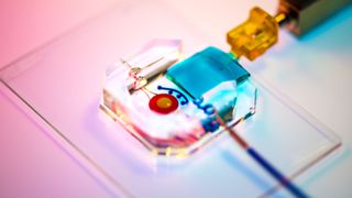
'Organ-on-chip' shows how uterus coaxes embryo to implant in early pregnancy
A tiny device replicates the interface between uterine tissue and the cells of the placenta in early pregnancy.

Scientists designed a tiny "organ-on-a-chip," about the size of a quarter, that replicates early pregnancy, when the embryo implants in the lining of the uterus.
In a report published earlier this year in the journal Nature Communications, the device's designers described the new technology in detail. The small device is made of clear silicone rubber, the same material used in some contact lenses, and has two chambers: one to hold placental cells and one for teensy, 3D blood vessels, according to Penn Medicine News. A barrier runs between the two chambers and mimics the uterine tissue that would run beneath an embryo implanted in the womb.
In experiments, the team used the device to observe how trophoblasts — cells that help the embryo attach to the uterus and later form part of the placenta — migrate towards uterine blood vessels prior to the embryo's implantation.
Cells lining the blood vessels actually aid trophoblasts on their journey by switching on specific genes and secreting various proteins, and with their new system, the researchers could watch this process unfold. The research also revealed that certain immune cells may play a key, but unsung, role in preparing the uterus for implantation.
"Our implantation-on-a-chip system was modeled to study the interactions between the mother and the placental tissue of the baby," said co-senior author Dr. Monica Mainigi, an associate professor of Obstetrics and Gynecology and the fellowship director of Reproductive Endocrinology and Infertility at the Hospital of the University of Pennsylvania.
"The reason we went after this question was because humans are fairly unique both in how the placenta attaches to the mother and in the interface between the maternal and placental cells," Dr. Mainigi told Penn Medicine News. "Now that we’ve shown the relevance of this system, we can look at the individual cell types and really get a much better understanding of what each individual cell type is doing."
Sign up for the Live Science daily newsletter now
Get the world’s most fascinating discoveries delivered straight to your inbox.

Nicoletta Lanese is the health channel editor at Live Science and was previously a news editor and staff writer at the site. She holds a graduate certificate in science communication from UC Santa Cruz and degrees in neuroscience and dance from the University of Florida. Her work has appeared in The Scientist, Science News, the Mercury News, Mongabay and Stanford Medicine Magazine, among other outlets. Based in NYC, she also remains heavily involved in dance and performs in local choreographers' work.
