It's quick and easy to access Live Science Plus, simply enter your email below. We'll send you a confirmation and sign you up for our daily newsletter, keeping you up to date with the latest science news.
(Image credit: Henrik Lauridsen and Kasper Hansen, MR Research Center, Aarhus University Hospital, Skejby, Denmark)
Fasting Burmese pythons were scanned before and at two, 16, 24, 40, 48, 72 and 132 hours after ingestion of one rat. The succession of images revealed a gradual disappearance of the body of the rat, accompanied by an overall expansion of the snake's intestine, shrinking of the gallbladder, and a 25-percent increase in heart volume.
Python Eating Three Rats
(Image credit: Henrik Lauridsen and Kasper Hansen, MR Research Center, Aarhus University Hospital, Skejby, Denmark)
In one experiment, scientists fed a python three rats and watched as the snacks descended through the snake's gut and vanished.
Python Eating One Rat
(Image credit: Henrik Lauridsen and Kasper Hansen, MR Research Center, Aarhus University Hospital, Skejby, Denmark)
The scientists used magnetic resonance imaging and computed tomography to scan a python's body while the snake was eating a rat, showing the rat mid-way through the body as it was being digested.
Henrik Lauridsen and Kasper Hansen, MR Research Center, Aarhus University Hospital, Skejby, Denmark
(Image credit: Henrik Lauridsen and Kasper Hansen, MR Research Center, Aarhus University Hospital, Skejby, Denmark)
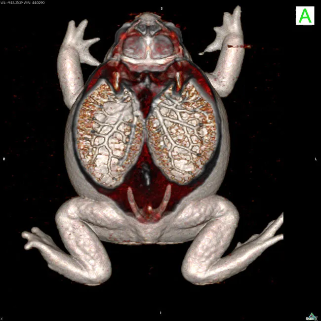
A live cane toad's lungs are captured in action with a computed tomography (CT) technique.
Turtle Blood Vessels
(Image credit: Henrik Lauridsen and Kasper Hansen, MR Research Center, Aarhus University Hospital, Skejby, Denmark)
A live turtle's blood vessels are captured in action with a magnetic resonance imaging technique.
Alligator Anatomy
(Image credit: Henrik Lauridsen and Kasper Hansen, MR Research Center, Aarhus University Hospital, Skejby, Denmark)
A CT scan reveals the skeletal anatomy of a live alligator.
Aligator MRI
(Image credit: MR Research Center, Aarhus University Hospital, Denmark)
Scientists used MRI scanning to reveal the inner anatomy of an alligator. Contrasting agents were used to highlight specific organs in the MRI and computed tomography (CT) images.
Sign up for the Live Science daily newsletter now
Get the world’s most fascinating discoveries delivered straight to your inbox.
Jeanna Bryner is managing editor of Scientific American. Previously she was editor in chief of Live Science and, prior to that, an editor at Scholastic's Science World magazine. Bryner has an English degree from Salisbury University, a master's degree in biogeochemistry and environmental sciences from the University of Maryland and a graduate science journalism degree from New York University. She has worked as a biologist in Florida, where she monitored wetlands and did field surveys for endangered species, including the gorgeous Florida Scrub Jay. She also received an ocean sciences journalism fellowship from the Woods Hole Oceanographic Institution. She is a firm believer that science is for everyone and that just about everything can be viewed through the lens of science.

 Live Science Plus