Baby's Rare Brain Tumor Had Teeth

A 4-month-old infant in Maryland may be the first person to have had teeth form in his brain as a result of a specific type of rare brain tumor, according to a new report of the case.
The boy is doing well now that his tumor has been removed, and doctors say the case sheds light on how these rare tumors develop.
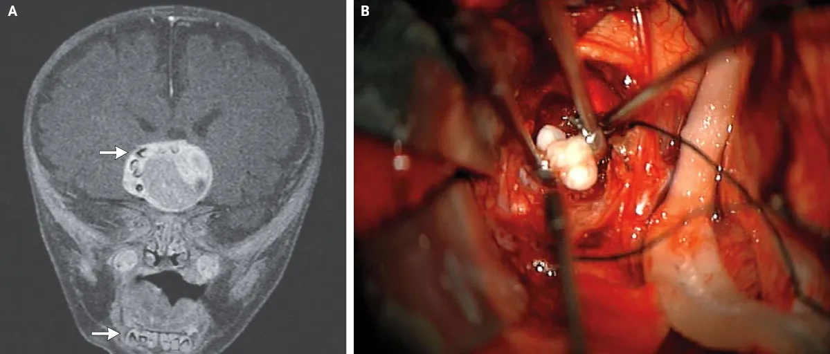
Doctors first suspected something might be wrong when the child's head appeared to be growing faster than is typical for children his age. A brain scan revealed a tumor containing structures that looked very similar to teeth normally found in the lower jaw.
Article continues belowThe child underwent brain surgery to have the tumor removed, during which doctors found that the tumor contained several fully formed teeth, according to the report. [14 Oddest Medical Cases]
After an analysis of tumor tissue, doctors determined the child had a craniopharyngioma, a rare brain tumor that can grow to be larger than a golf ball, but does not spread.
Researchers had always suspected that these tumors form from the same cells involved in making teeth, but until now, doctors had never seen actual teeth in these tumors, said Dr. Narlin Beaty, a neurosurgeon at the University of Maryland Medical Center, who performed the boy's surgery along with his colleague, Dr. Edward Ahn, of Johns Hopkins Children's Center.
"It's not every day you see teeth in any type of tumor in the brain. In a craniopharyngioma, it's unheard of," Beaty said.
Get the world’s most fascinating discoveries delivered straight to your inbox.
Craniopharyngiomas commonly contain calcium deposits, "but when we pulled out a full tooth...I think that’s something slightly different," Beaty told Live Science.
Teeth have been found in people's brains before, but only in tumors known as teratomas, which are unique among tumors because they contain all three of the tissue types found in an early-stage human embryo, Beaty said. In contrast, craniopharyngiomas have only one layer of tissue.
The boy's case provides more evidence that craniopharyngiomas do indeed develop from the cells that make teeth, Beaty said.
These tumors are most often diagnosed in children ages 5 to 14, and are rare in children younger than 2, according to the National Cancer Institute.
The boy is progressing well in his development, the researchers said. However, because craniopharyngiomas are tumors of the pituitary gland — a gland in the brain that releases many important hormones — they often cause hormone problems.
In the boy's case, the tumor destroyed the normal connections in the brain that would allow certain hormones to be released, Beaty said, so he will need to receive hormone treatments for the rest of his life to replace these hormones, Beaty said.
"He's doing extremely well, all things considered," Beaty said. "This was a big tumor right in the center of his brain. Before the modern surgical era this child would not have survived," Beaty said.
The teeth were sent to a pathologist for further study, Beaty said, and generally, these types of tissue samples are saved for many years in case more investigation is needed.
The report is published in the Feb. 27 issue of the New England Journal of Medicine.
Follow Rachael Rettner @RachaelRettner. Follow Live Science @livescience, Facebook & Google+. Original article on Live Science.

Rachael is a Live Science contributor, and was a former channel editor and senior writer for Live Science between 2010 and 2022. She has a master's degree in journalism from New York University's Science, Health and Environmental Reporting Program. She also holds a B.S. in molecular biology and an M.S. in biology from the University of California, San Diego. Her work has appeared in Scienceline, The Washington Post and Scientific American.