
 Live Science Plus


Code of Life: Photos of DNA Structures

From human genomes
G-quadruplex inspired

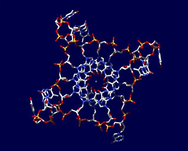
This image is based on an x-ray crystal structure of a G-quadruplex formed from the DNA sequence found in human genomes.
X-ray crystal structure

This image is based on an x-ray crystal structure of a G-quadruplex formed from the DNA sequence found in human genomes.

From start to finish

This graph shows the formation of DNA G-quadruplex structures. DNA can adopt structures that are alternative to the double-helix and can be visualised in human cancer cells by microscopy, thanks to an antibody that target them.
Cancer cells

This image shows, on the left, DNA G-quadruplex structures (red foci) are present in human cancer cell nuclei (blue). On the right, more structures are visualized after treatment with a G-quadruplex-stabilising small molecule compared to the untreated control on left.
Chromosomes

In this image, DNA G-quadruplex structures (red foci) can be visualised in human chromosomes (blue) and are present both at telomeres (arrow), areas at the end of chromosomes, but also throughout the chromosomes.
Telomeres

In this image DNA G-quadruplex structures (red foci) can be visualised in human chromosomes (blue) and are present both at telomeres (arrow) and throughout the chromosomes. Scale bar corresponds to 2.5 um.
Human chromosomes

In this image DNA G-quadruplex structures (red foci) can be visualised in human chromosomes (blue) and are present both at telomeres (arrow) and throughout the chromosomes. Scale bar corresponds to 2.5 um.
Representation of quadraplex

Professor Shankar Balasubramanian in front of a mural in his office in Cambridge's Department of Chemistry. Painting is representation of quadruplex DNA called 'Living by the code' by artist Annie Newman.
Living by the code

A mural on the wall of Professor Shankar Balasubramanian's office in Cambridge's Department of Chemistry. Painting is representation of quadruplex DNA called 'Living by the code' by artist Annie Newman.
