For the first time, scientists have reconstructed a three-dimensional circuit of connected cells in the brain's seat of consciousness. Their new approach, which involves the use of high-tech microscopes and a supercomputer, offers the unprecedented opportunity to unravel the complex wiring of the brain by navigating through the tangled and dense jungle of cells — similar to the way Google crawls the Web.
The research, published by two separate teams in the March 10 issue of the journal Nature, demonstrates the possibility of tackling questions about brain function that traditional methods can't address. One study was led by neurobiologist Clay Reid of Harvard University, and the other was spearheaded by Winfried Denk at the Max Planck Institute for Medical Research in Heidelberg, Germany. [Image of brain-cell map]
As brain-imaging techniques advance, scientists have had great success looking at the activity of brain cells. While this answers the "what are they doing" question, it hasn't shed light on the "how are they doing it" mystery.
Latest Videos From
So the researchers turned to the cerebral cortex, the outside layer of the brain implicated in higher-order mental functions, including memory.
"Cortical circuits are very big, and so far we've been looking at networks of cells wired two cells at a time, or a handful of connections at a time," Reid told LiveScience. "This combination of techniques gives us the hope that in the coming decade we'll be able to look and see the physiology of literally every cell in a local network."
The individual techniques Reid used are not new, but he and his team developed painstaking procedures for matching brain-structure data with neural recordings to recreate a circuit in the visual system of mice.
They first had lab mice view lit-up bars on a screen as they measured the activity of about a dozen neurons known to play a role in mouse vision.
To figure out how these neurons were physically connected into a circuit, the researchers then turned to the electron microscope (EM), which produced high-resolution images of the animals' brain tissues by beaming electrons onto more than 1,200 tiny, adjacent slices of the brain.
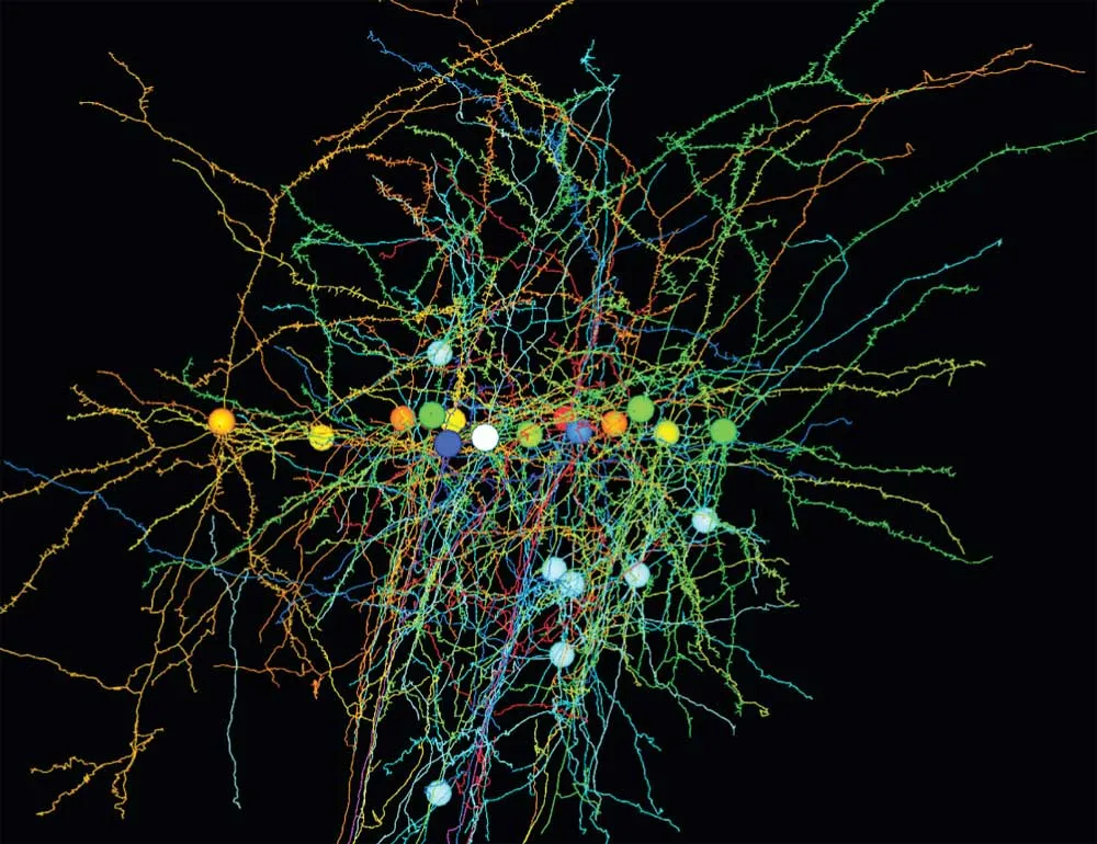
They used a supercomputer to stitch together millions of high-resolution images, resulting in a three-dimensional map that looked like a forest of indecipherable wires, Reid said.
To locate the data of interest within the microscope images, the researchers manually traced the neurons they had already recorded and mapped out hundreds of their connections with nearby cells.
They focused on 10 brain cells that seemed to be critical to vision in the mice. "They spent three months of their lives drawing three-dimensional stick figures of the 10 neurons," Reid said. They essentially crawled through the brain's dense thicket, jumping from neuron to neuron to create a partial diagram of the mouse brain's visual circuit, helping to answer the question, "How does the brain see?" Reid said. [Effort to Map Human Brain Faces Complex Challenges]
Recent progress in data collection, storage and processing made the research possible, and further advances will allow scientists to probe circuits of hundreds or thousands of neurons, Reid said. "That's when it really will get interesting: when we have a much bigger and more densely connected network."
“This study is not the last word,” Reid added. “It’s very much the first attempt at something that’s very exciting that we hope will give a lot of answers in the coming years.”