 Live Science Plus


You are now subscribed
Your newsletter sign-up was successful
Want to add more newsletters?

Delivered Daily
Daily Newsletter
Sign up for the latest discoveries, groundbreaking research and fascinating breakthroughs that impact you and the wider world direct to your inbox.

Once a week
Life's Little Mysteries
Feed your curiosity with an exclusive mystery every week, solved with science and delivered direct to your inbox before it's seen anywhere else.

Once a week
How It Works
Sign up to our free science & technology newsletter for your weekly fix of fascinating articles, quick quizzes, amazing images, and more

Delivered daily
Space.com Newsletter
Breaking space news, the latest updates on rocket launches, skywatching events and more!

Once a month
Watch This Space
Sign up to our monthly entertainment newsletter to keep up with all our coverage of the latest sci-fi and space movies, tv shows, games and books.

Once a week
Night Sky This Week
Discover this week's must-see night sky events, moon phases, and stunning astrophotos. Sign up for our skywatching newsletter and explore the universe with us!
Join the club
Get full access to premium articles, exclusive features and a growing list of member rewards.

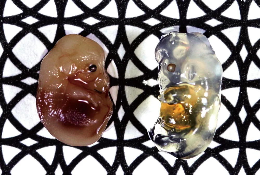
Scientists are seeing deeper into the brain than ever before with the help of a new technique that allows them to turn tissues transparent.
"Our current experiments are focused on the mouse brain, but applications are neither limited to mice, nor to the brain," study researcher Atsushi Miyawaki, of RIKEN Brain Science Institute in Japan, said in a statement. "We envision using Scale on other organs such as the heart, muscles and kidneys, and on tissues from primate and human biopsy samples."
Visualizing tissues

Unlike previous techniques to make tissues transparent, Scale, which uses a simple liquid, doesn't interfere with the fluorescent dyes scientists use to highlight certain tissues.
"More and more researchers are interested in obtaining large-scale subcellular resolution 3-D reconstructions of the fluorescent structures," Miyawaki told LiveScience in an email. "The Scale technique renders biological specimens transparent while preserving fluorescent signals, and is thus very useful."
By labeling specific types of cells with different fluorescent colors, researchers can see how they interact inside tissues. The Scale technique can be used in concert with the "brainbow" labeling developed in 2007, which labels brain cells with 90 different colors; the two techniques would visualize how different kinds of brain cells interact in three dimensions, instead of two.
The treatment also enables researchers to see deep into tissues — up to 0.15 inches (4 millimeters) into the brain — a distance that is limited only by current microscopes' ability to "see" at different depths, which the researchers hope will be improved in the near future.

Transparent future
They are currently studying the anatomical differences between different areas of the mouse brain. They are also working on developing a similar technique that could be used on living samples, though this one wouldn't reach nearly as far into tissues.
"We are currently investigating another, milder candidate reagent, which would allow us to study live tissue in the same way, at somewhat lower levels of transparency," Miyawaki said. "This would open the door to experiments that have simply never been possible before."
The study was published Aug. 30 in the journal Nature Neuroscience.
You can follow LiveScience staff writer Jennifer Welsh on Twitter @microbelover. Follow LiveScience for the latest in science news and discoveries on Twitter @livescience and on Facebook.

