 Live Science Plus


Using lasers, scientists can now surgically blast holes thinner than a human hair in the heads of live fruit flies, allowing researchers to see how the flies' brains work.
The researchers also successfully tested this technique on worms, ants and mice.
Microscopically peering into living animals can help scientists learn more about key details of these animals' biology. For instance, tiny glass windows surgically implanted into the sides of living mice can help researchers study how cancers develop in real time and evaluate the effectiveness of potential medicines.
Article continues belowSurgically preparing small live animals for such "intravital microscopy" is often time-consuming and requires considerable skill and dexterity. Now, Supriyo Sinha, a systems engineer at Stanford University in California, and his colleagues have developed a way to prepare live animals for such microscopy that is both fast — taking less than a second — and largely automated.
Fruit-fly brains
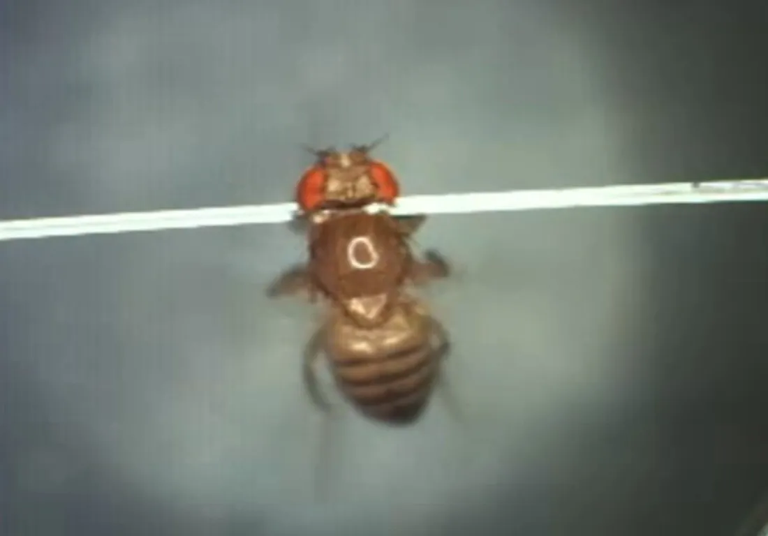
To conduct this procedure, scientists first cooled fruit flies to anesthetize them. Then, the researchers carefully picked up the insects with tweezers and glued them to the tops of glass fibers in order to immobilize the flies' bodies and heads. Then, using a high-energy pulsed ultraviolet laser, the researchers blasted holes measuring 12 to 350 microns wide in the flies' heads. (In comparison, the average human hair is about 100 microns wide.) They then applied a saline solution to exposed tissue to help keep the fly brains healthy. [See Experiment Video and Images of Fruit Fly's Brain]
Using lasers enabled the researchers to create these "windows" up to 100 times faster than they could be created manually. Moreover, these laser-cut windows were apparently substantially gentler on fly health than ones created by conventional surgery — the researchers could image brain activity for longer than they could using the conventional method, up to 18 hours, about five to 20 times longer than prior microscopy studies of living, hand-dissected flies.

"The induced trauma to the fly is minimized, and the fly can remain alive longer," Sinha told LiveScience. "Learning and memory experiments in which the brain is imaged before and after training is possible."
Prior research had tried using laser surgery to open holes in animals for intravital microscopy before. Compared to past work that used infrared, visible or larger-wavelength ultraviolet lasers, this new technique can remove tissue more quickly or cause less collateral damage in the brain.
Sinha and his colleagues also successfully tested their technique on anesthetized and immobilized ants, nematode worms and mice. "Our main motivation is to better understand neural circuits, and faster screening and imaging could better help us reverse-engineer these circuits," Sinha said.
From one to 100
The scientists are also developing to automatically capture, mount and align the insects for laser surgery. Their short-term goal is to build a system that can hold a dozen flies.
"We are trying to streamline the procedure such that the experimentalist only has to press one button to have the system pick and mount and align 12 flies; a second button that would surgically remove the cuticle and apply saline to the 12 flies; and a third button to start imaging the 12 flies under predetermined stimulation," Sinha said.
Ultimately the researchers would like to simultaneously image the brains of about 100 awake fruit flies with a push of a few buttons, Sinha added.
"Our goal is to have this arrayed imaging technology adopted by a few other labs in the world," Sinha said. "These imaging centers could be used by fly biologists all over the world to conduct new classes of experiments that would not be possible or would be too impractical using traditional techniques."
The scientists detailed their findings online Oct. 28 in the journal Proceedings of the National Academy of Sciences.
Follow us @livescience, Facebook & Google+. Original article on LiveScience.

