
 Live Science Plus


Glowing Green Orbs & Pink Butterflies Revealed in Winning Bio-Art Images

A pink butterfly, fluorescent mountains and glowing green orbs surrounded by bubbles are some of the imagery that appears in the winning entries in a "bio-art" competition, which sought to highlight the most artistic portrayals of biomedical research.
This year, 10 images out of about 100 entries were honored in the first Bio-Art Competition, created by the Federation for American Societies for Experimental Biology (FASEB). The winning entries weren't ranked and all were generated by scientists as a byproduct of of biomedical research.
Art wasn't the purpose of research to create artificial stem cell factories, explore the biological basis for psychiatric disease or look at the production of new neurons in the adult brain. Even so, the winning entries included brightly colored and sometimes abstract images.

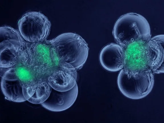
The winners include one image depicting a scaffold, which resembles the weave of a fabric, upon which cells can grow to form new tissue. Glowing green orbs surrounded by bubbles reveal systems intended to produce muscle stem cells; and images of species of electric fish are trailed by recordings of the discharges.
"Electric fish recognize other members of their own species using the species-specific waveforms of these heartbeatlike discharges," write the team lead by Matthew Arnegard of the Fred Hutchinson Cancer Research Center in Seattle who created the image. (Under the contest guidelines, the image and the statement accompanying it should be visually arresting and clearly communicate a cutting-edge concept in biomedical science.)

Another winning entry depicts what looks like a fluorescent three-dimensional map, but is actually genetically labeled cells covering the walls of capillaries in a mouse kidney. [See Photos of the Winning Bio-Art]
University of Iowa's Li-Hsien Lin captured an image that appears to be a pink butterfly, but is actually a rat spinal cord showing the distribution of different types of enzymes. Understanding how these enzymes work and interact in the nervous system, Lin writes, could lead to better treatments for cardiovascular diseases such as hypertension and heart failure.

Other honorees included: abstract art created from tissue from a colon biopsy stained for a particular receptor; converging fibers that form the optic nerve in a mouse retina and their attendant immune cells; a 3D image of the limb from a transgenic, embryonic mouse, with bright colors differentiating the muscles, tendons, bones and nerves; and the growth of new neurons in the adult brain.
FASEB is a coalition of biomedical research associations in the United States. For this competition, the organization sought original, laboratory-based images produced by current or former National Institutes of Health-funded investigators, contractors, trainees or members of FASEB constituent societies, according to a news release.
You can follow LiveScience senior writer Wynne Parry on Twitter @Wynne_Parry. Follow LiveScience for the latest in science news and discoveries on Twitter @livescience and on Facebook.
