
 Live Science Plus


The Human Skeletal System
Reference Article: Facts about the human skeletal system, its function and common skeletal diseases.
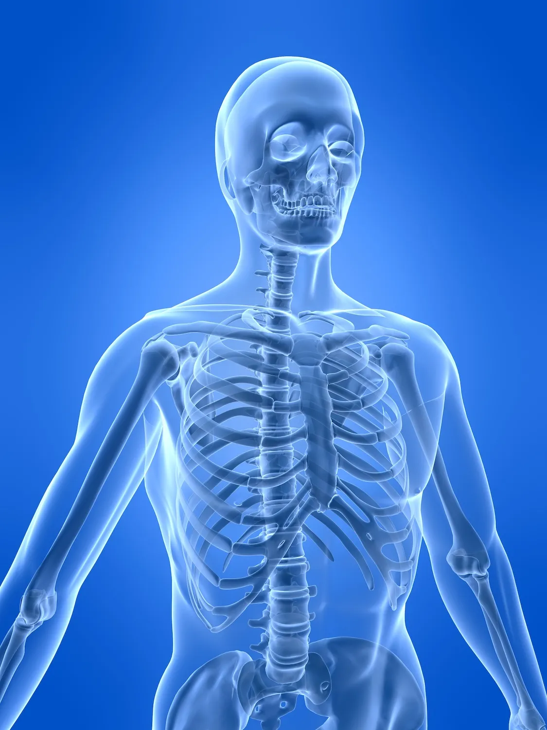
The human skeletal system is not quite as simple as the popular children's song suggests. The "head bone" (actually made up of 22 separate bones) is not connected to the "neck bone," but rather to a series of small bones that go all the way down the back. And the "toe bone" is actually made up of several bones that connect to another set of bones that provide structure for the foot. In total, the human skeleton consists of a whopping 206 bones.
In addition to all those bones, the human skeletal system includes a network of tendons, ligaments and cartilage that connect the bones together. The skeletal system provides the structural support for the human body and protects our organs. Our bones also serve several other vital functions, including producing blood cells and storing and releasing fats and minerals, according to the National Center for Biotechnology Information (NCBI).
Development and structure of the skeleton
Infants are born with about 300 separate bones, according to Nemours, a nonprofit children's health provider. As a child grows, some of those bones fuse together until growth stops, typically by the age of 25, leaving the skeleton with 206 bones.
Our bones are separated into two categories based on the purpose and location of the bones: The axial skeleton and the appendicular skeleton, according to the SEER program of the National Cancer Institute.
The axial skeleton contains 80 bones, including the skull, spine and rib cage. It forms the central structure of the skeleton, with the function of protecting the brain, spinal cord, heart and lungs.
The remaining 126 bones make up the appendicular skeleton; they include the arms, legs, shoulder girdle and pelvic girdle. The lower portion of the appendicular skeleton protects the major organs associated with digestion and reproduction and provides stability when a person is walking or running. The upper portion allows for a greater range of motion when lifting and carrying objects.
Bones are further classified by their shape: long, short, flat, irregular or sesamoid, according to SEER.
Get the world’s most fascinating discoveries delivered straight to your inbox.
- Long bones are found in the arms, legs, fingers and toes. These bones are longer than they are wide and are cylindrical. They move when the muscles around them contract, and they are the most mobile parts of the skeleton.
- Short bones are found in the wrists and ankles and are about equal in their length, width and thickness.
- Flat bones make up the skull, shoulder blades, sternum and ribs. These curved, thin bones protect internal organs and provide an anchor for muscles.
- Irregular bones are those in the spinal cord and face, which, because of their unique dimension, don't fit in any of the other shape categories.
- Sesamoid bones are found in the hands, wrists, feet, ears and knees. These small, round bones are embedded in tendons and protect them from the great pressure and force they encounter.
There are some variations between male and female skeletons. For example, the female pelvis is typically more broad, thin, and round than the male pelvis, according to the National Museum of Natural History. [Image Gallery: The BioDigital Human]
What's inside your bones?

All about your body's skeleton, the framework of bones that keeps you together.
Three main types of material make up every bone in your body: compact bone, spongy bone and bone marrow, according to the School of Life Sciences at Arizona State University.
Approximately 80% of every bone is compact bone, which is the hardest and strongest type of bone and is what allows the body to support its weight. Compact bone makes up the outer layers of the bone and protects the inner parts of the bones where many vital functions occur, such as bone marrow production. Compact bone consists primarily of cells called osteocytes. Microscopic passages in between the cells to allow nerves and blood vessels to pass through.
About 20% of each bone is spongy bone, which is filled with large holes and passages. Most often found toward the ends of individual bones, the spongy bone material is filled with bone marrow, nerves and blood vessels.
Two types of bone marrow fill the pores in spongy bone. Approximately half is red bone marrow, which is found mainly within flat bones such as shoulder blades and ribs. This is where all red and white blood cells and platelets (cells that help a cut stop bleeding) are made. Infant's bones contain all red bone marrow to produce enough blood cells to keep up with the youngsters' growth.
The other half of marrow is yellow bone marrow, which is found in long bones, such as thigh bones, and consists primarily of fat. Blood vessels run through both types of bone marrow to deliver nutrients and remove waste from the bones.
There are four main types of cells within bones: Osteoblasts, osteocytes, osteoclasts and lining cells, according to the NCBI.
Osteoblasts are cells that create new or repair existing bone material as the bones grow or break. The cells create a flexible material called osteoid and then fortify it with minerals to harden and strengthen it. When osteoblasts successfully finish their job, they retire to become osteocytes or lining cells.
Osteocytes, found in the compact bone, are responsible for exchanging minerals and communicating with other cells in the vicinity. They are formed from old osteoblasts that have gotten stuck in the center of bones.
Osteoclasts break down existing bone material and reabsorb it. These cells often work with osteoblasts to heal and reshape bone after a break (the osteoclasts break down the extra callus formed by the healing process) to make room for new blood vessels and nerves and to make bones thicker and stronger.
Lining cells are flat bone cells that completely cover the outside surface of bones. Their primary function is controlling the movement of minerals, cells and other materials into and out of the bones.
Diseases of the skeletal system
As with any part of the human body, bones are susceptible to injury and disease.
Some of the most common diseases that can affect the skeletal system include:
- Osteoporosis is a disease that causes the density and strength of bones to decrease because bone loss occurs faster than bone growth. It can be caused by genetics or unhealthy lifestyle habits (such as lack of calcium or vitamin D, and heavy smoking or drinking with little exercise).
- Leukemia is a type of cancer that starts in the bone marrow and the lymphatic system, according to the Mayo Clinic. Several types of leukemia affect various blood cells and other systems of the body.
- Osteoarthritis is a disease that causes the breakdown of the cartilage that protects the ends of bones in joints. This lack of cartilage leads to bone-on-bone rubbing, which can cause significant pain, damage to the bones and connective tissues, inflammation of the surrounding tissue and restricted motion, according to the Mayo Clinic.
Additional resources:
- Learn more about the skeletal structure and function from Khan Academy.
- Check out some pictures of cool animal skeletons from the Oxford University Museum of Natural History.
- Learn more about the differences between the male and female skeleton, from the Smithsonian Institution.
This article was updated on Oct. 18, 2021, by Live Science contributor Ben Biggs.
This article is for informational purposes only, and is not meant to offer medical advice.
Kim Ann Zimmermann is a contributor to Live Science and sister site Space.com, writing mainly evergreen reference articles that provide background on myriad scientific topics, from astronauts to climate, and from culture to medicine. Her work can also be found in Business News Daily and KM World. She holds a bachelor’s degree in communications from Glassboro State College (now known as Rowan University) in New Jersey.