


NASA Imaging Technology Helps Fight Breast Cancer
The same software used by NASA scientists to determine the depths of lakes from space could also be used by doctors to detect changes in breast density during mammograms.
The imaging technology was approved in July by the U.S. Food and Drug Administration, under the name MED-SEG, for use in medical reports, though the software can't be used for diagnosis because clinical tests haven't been conducted yet.
A big problem with mammography is that it's hard to detect cancers in a woman's breasts if the tissue is too dense, said Dr. Molly Brewer, a professor of gynecologic oncology at the University of Connecticut Health Center. That could lead to missed opportunities to find breast cancer early.
"What happens when a radiologist reads a mammogram, CT scan or an MRI, is they look at differences in density, but that's subject to the human eye," Brewer said at a news conference today. "That's where differences can occur we may not see with our eyes what a computer can see."
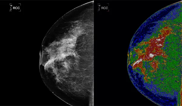
[Images: See pictures of dense tissue using MED-SEG and using a traditional mammogram ]
How it works
The MED-SEG could reduce the importance of subjectivity in reading mammograms, and allow doctors to get clearer results from an imaging test. The software may also fill a void between mammograms, which don't always detect changes in density, and magnetic resonance imaging tests (MRIs), which are more sensitive but also expensive, and can falsely reveal problems that aren't really there, Brewer said.
Get the world’s most fascinating discoveries delivered straight to your inbox.
Accurately measuring breast density is important because previous research has found that among women with early breast cancer, those with the highest breast density are at the highest risk for cancer recurrence .
And mammograms currently miss up to 20 percent of breast cancers . Doctors have a harder time diagnosing women with dense breast tissue because the tissue looks similar to tumors in mammograms, according to the National Cancer Institute.
Brewer is working with Bartron Medical Imaging Inc., the owner of MED-SEG, to develop clinical trials to test the software in doctors' offices. The trials are set to start within the next six to eight months.
The software works because it doesn't look only at an image's individual pixels, which don't provide much information or context by themselves, said developer James C. Tilton, a computer engineer at NASA's Goddard Space Flight Center in Maryland, who developed the software.
Instead, it groups pixels based on their level of detail, and distinguishes hard-to-see details in the image, he said.
Detailed images revealed
"[I] was surprised that something I developed for a large-scale earth science study could be applied effectively on such a small scale," Tilton said.
For example, in a satellite image of the Earth, all lakes would appear blue and all land would appear green. But in an image using the software, shallow lakes would have a different shade of blue than deeper lakes, he said.
The same goes for images of breast cells. Without the software, a cell is hard to distinguish from its background. But the software enhances the activity happening in the cell, making it easier to see fine detail, Tilton said.
The technology also has potential use in examining vegetation for agricultural purposes, said Nona Cheeks, chief of the Innovative Partnerships Program Office at NASA.
- 3 Lifestyle Choices Lower Breast Cancer Risk, Regardless of Family History
- The 10 Deadliest Cancers and Why There's No Cure
- Breast Cancer: Symptoms, Treatments and Prevention
