In Images: 2012 Nikon Small World Contest Winners
Get the world’s most fascinating discoveries delivered straight to your inbox.
You are now subscribed
Your newsletter sign-up was successful
Want to add more newsletters?

Delivered Daily
Daily Newsletter
Sign up for the latest discoveries, groundbreaking research and fascinating breakthroughs that impact you and the wider world direct to your inbox.

Once a week
Life's Little Mysteries
Feed your curiosity with an exclusive mystery every week, solved with science and delivered direct to your inbox before it's seen anywhere else.

Once a week
How It Works
Sign up to our free science & technology newsletter for your weekly fix of fascinating articles, quick quizzes, amazing images, and more

Delivered daily
Space.com Newsletter
Breaking space news, the latest updates on rocket launches, skywatching events and more!

Once a month
Watch This Space
Sign up to our monthly entertainment newsletter to keep up with all our coverage of the latest sci-fi and space movies, tv shows, games and books.

Once a week
Night Sky This Week
Discover this week's must-see night sky events, moon phases, and stunning astrophotos. Sign up for our skywatching newsletter and explore the universe with us!
Join the club
Get full access to premium articles, exclusive features and a growing list of member rewards.
Holiday lights
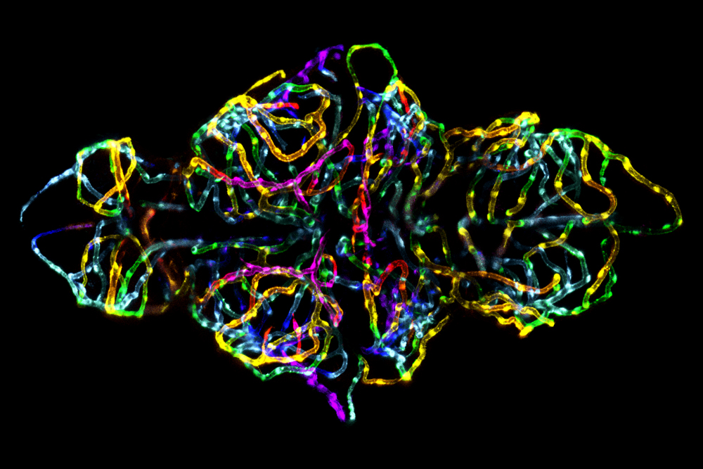
The blood-brain barrier in a live zebrafish embryo captured using the Confocal technique at 20 times magnification.
Adorable arachnids

Live newborn lynx spiderlings shot using Reflected Light, Figer Optics and Image Stacking Techniques at six times magnification.
Up close and personal
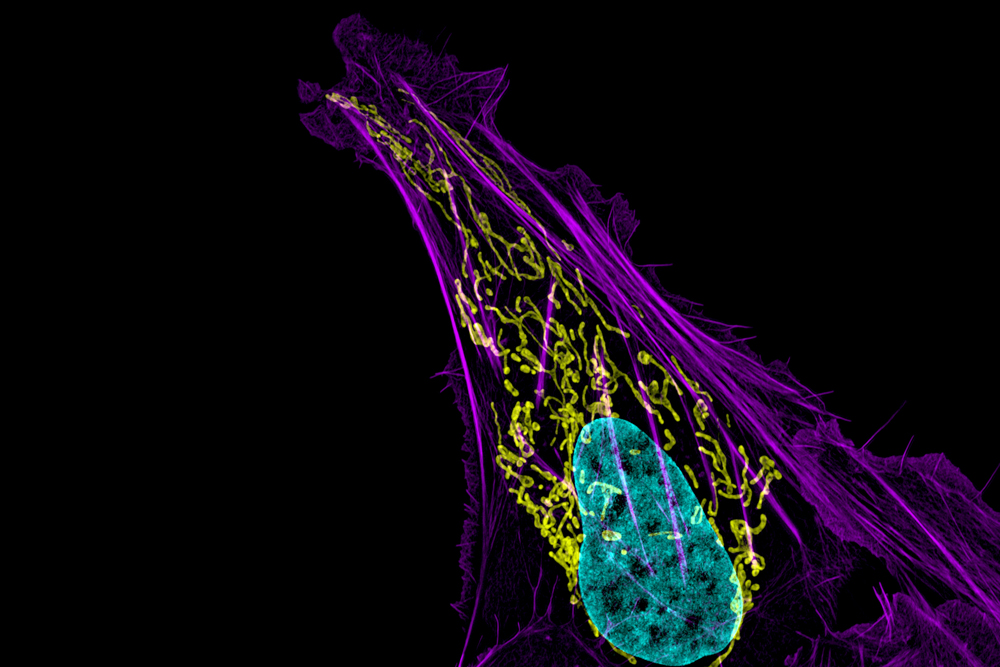
Human bone cancer (osteosarcoma) showing actin filaments (purple), mitochondria (yellow), and DNA (blue) captured with the Structured Illumination Microsopy (SIM) technique at 63 times magnification.
In development
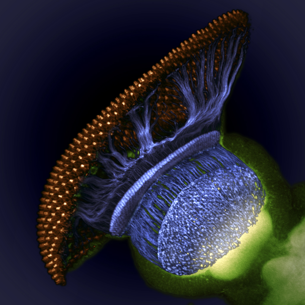
Drosophila melanogaster (fruit fly) visual system halfway through pupal development, showing retina (gold), photoreceptor axons (blue), and brain (green) imaged using the Confocal technique at 1500 times magnification.
Sparkling fluff
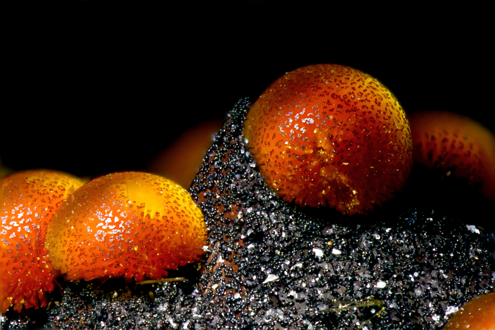
Cacoxenite (mineral) from La Paloma Mine, Spain in the Transmitted Light technique at 18 times magnification.
Coming in for a landing
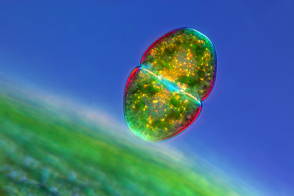
Cosmarium sp. (desmid) near a Sphagnum sp. leaf in the Polarized Light technique at 100 times magnification.
Heart-shaped
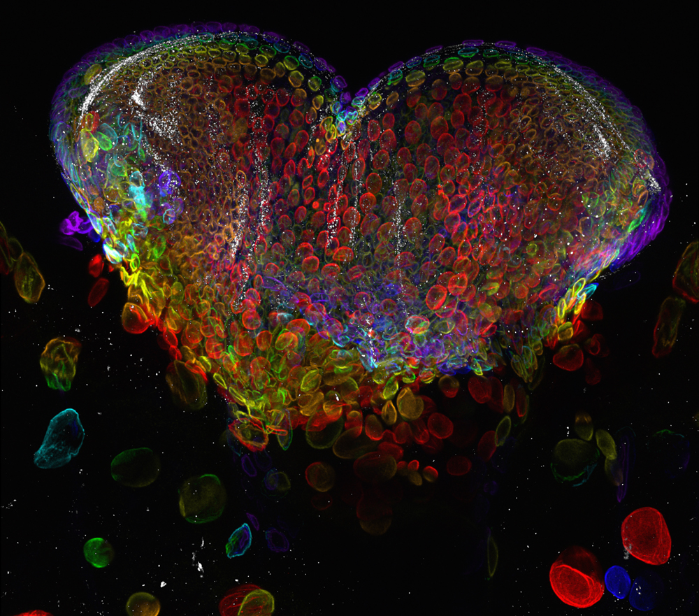
Eye organ of a Drosophila melanogaster (fruit fly) third-instar larvae pictured in the Confocal technique at 60 times magnification.
Get the world’s most fascinating discoveries delivered straight to your inbox.
Hairy legs
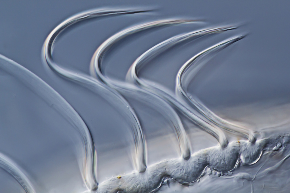
Pleurobrachia sp. (sea gooseberry) larva in the Differential Interference Contrast technique at 500 times magnification.
Family time
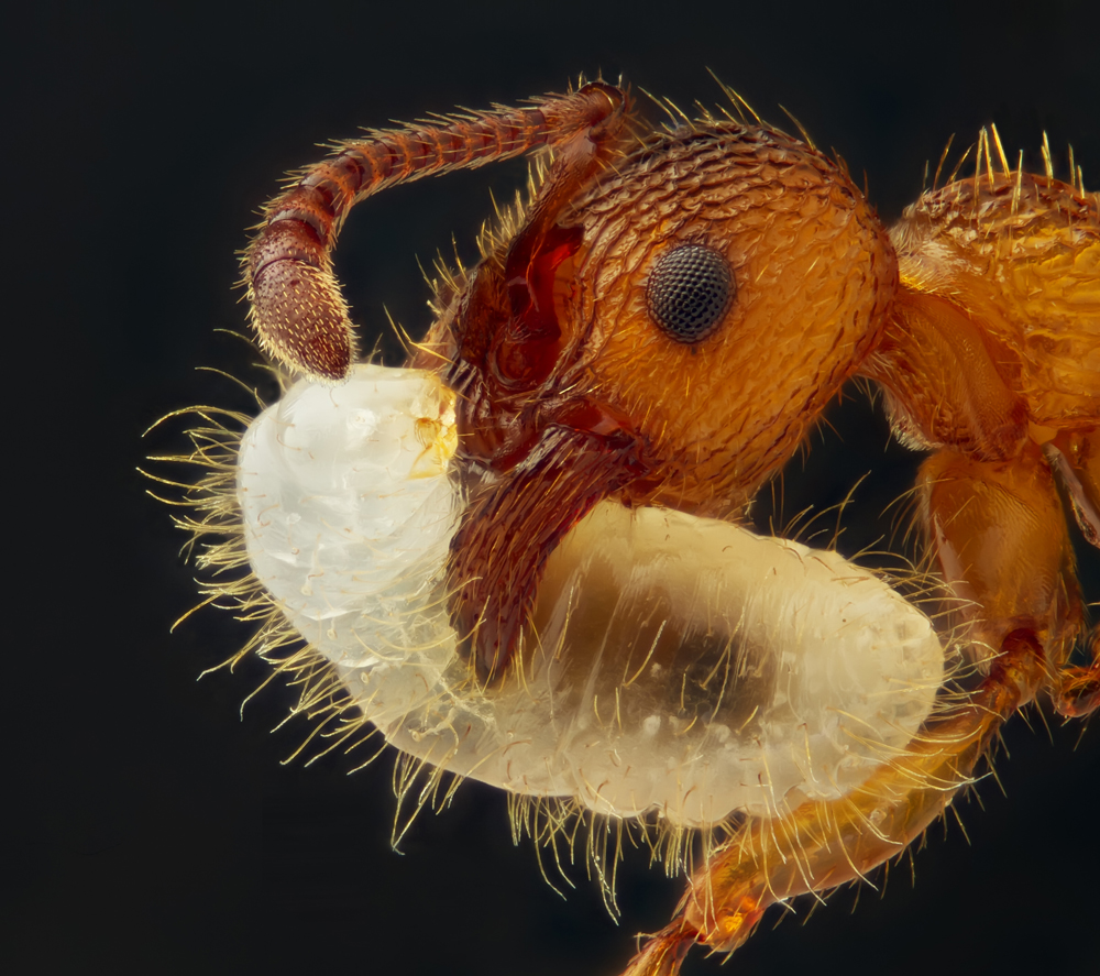
Myrmica sp. (ant) carrying its larva captured using Reflected Light and Image Stacking Techniques at five times magnification.
Spiny stars
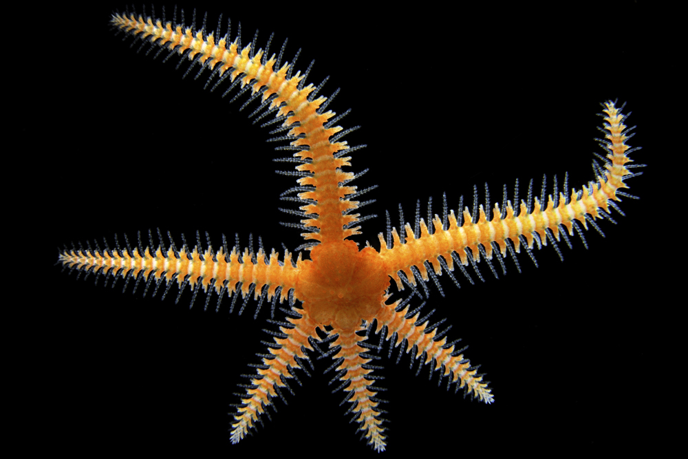
A brittle star imaged using Stereomicroscopy and Darkfield techniques at eight times magnification.
Electric lights
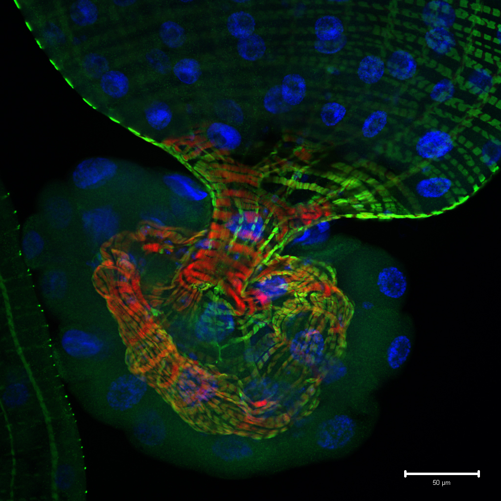
Single optical section through the tip of the gut of a Drosophila melanogaster larva expressing a reporter for Notch signaling pathway activity (green), and stained with cytoskeletal (red) and nuclear (blue) markers captured using the Confocal technique at 25 times magnification.
 Live Science Plus


