It's quick and easy to access Live Science Plus, simply enter your email below. We'll send you a confirmation and sign you up for our daily newsletter, keeping you up to date with the latest science news.
3D lymphangiogenesis assay. Cells sprout from dextran beads embedded in fibrin gel using the Fluorescence and Confocal techniques at 200 times magnification.
Miniscule worlds
(Image credit: Dr. Diana Lipscomb | George Washington University Department of Biological Sciences)
Sonderia sp. (a ciliate that preys upon various algae, diatoms, and cyanobacteria) pictured using the Nomarski Interference Contrast technique at 400 times magnification.
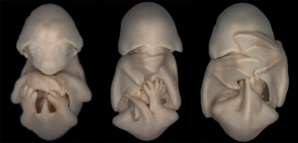
Tons of teeth
(Image credit: José R. Almodóvar Rivera | University of Puerto Rico Mayaguez Campus, Biology Department)
Pistil of Adenium obesum using the Image Stacking technique at 10 times magnification.
(Image credit: Andrea Genre | University of Turin Department of Life Sciences and Systems Biology)
Section of a Coccinella (ladybug) leg captured using the Confocal technique at 10 times magnification.
Swirls and twirls
(Image credit: Douglas Moore | University of Wisconsin - Stevens Point University Relations & Communications/Geology)
Fossilized Turitella agate containing Elimia tenera (freshwater snails) and ostracods (seed shrimp) captured using Stereomicroscopy at seven times magnification.
For the science geek in everyone, Live Science offers a fascinating window into the natural and technological world, delivering comprehensive and compelling news and analysis on everything from dinosaur discoveries, archaeological finds and amazing animals to health, innovation and wearable technology. We aim to empower and inspire our readers with the tools needed to understand the world and appreciate its everyday awe.

 Live Science Plus