Who Invented the Microscope?

For millennia, the smallest thing humans could see was about as wide as a human hair. When the microscope was invented around 1590, suddenly we saw a new world of living things in our water, in our food and under our nose.
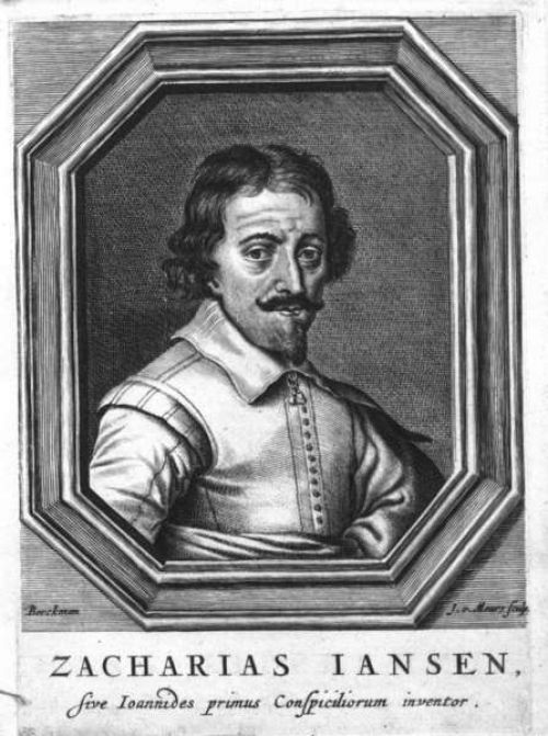
But it's unclear who invented the microscope. Some historians say it was Hans Lippershey, most famous for filing the first patent for a telescope. Other evidence points to Hans and Zacharias Janssen, a father-son team of spectacle makers living in the same town as Lippershey.
Janssen or Lippershey?
Hans Lippershey, also spelled Lipperhey, was born in Wesel, Germany in 1570, but moved to Holland, which was then enjoying a period of innovation in art and science called the Dutch Golden Age. Lippershey settled in Middelburg, where he made spectacles, binoculars and some of the earliest microscopes and telescopes.
Also living in Middelburg were Hans and Zacharias Janssen. Historians attribute the invention of the microscope to the Janssens, thanks to letters by the Dutch diplomat William Boreel.
In the 1650s, Boreel wrote a letter to the physician of the French king in which he described the microscope. In his letter, Boreel said Zacharias Janssen started writing to him about a microscope in the early 1590s, although Boreel only saw a microscope himself years later. Some historians argue Hans Janssen helped build the microscope, as Zacharias was a teenager in the 1590s.
Early microscopes
The early Janssen microscopes were compound microscopes, which use at least two lenses. The objective lens is positioned close to the object and produces an image that is picked up and magnified further by the second lens, called the eyepiece.
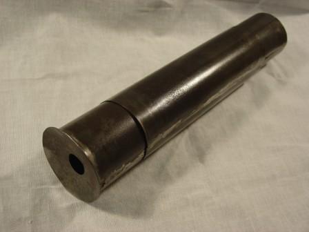
A Middelburg museum has one of the earliest Janssen microscopes, dated to 1595. It had three sliding tubes for different lenses, no tripod and was capable of magnifying three to nine times the true size. News about the microscopes spread quickly across Europe.
Get the world’s most fascinating discoveries delivered straight to your inbox.
Galileo Galilei soon improved upon the compound microscope design in 1609. Galileo called his device an occhiolino, or "little eye."
English scientist Robert Hooke improved the microscope, too, and explored the structure of snowflakes, fleas, lice and plants. He coined the term "cell" from the Latin cella, which means "small room," because he compared the cells he saw in cork to the small rooms that monks lived in. In 1665, and detailed his observations in the book "Micrographia."
Early compound microscopes provided more magnification than single lens microscopes; however, they also distorted the image more. Dutch scientist Antoine van Leeuwenhoek designed high-powered single lens microscopes in the 1670s. With these he was the first to describe sperm (or spermatozoa) from dogs and humans. He also studied yeast, red blood cells, bacteria from the mouth and protozoa. Van Leeuwenhoek's single lens microscopes could magnify up to 270 times larger than actual size. Single lens microscopes remained popular well into the 1830s, as all types of microscopes improved.
Scientists were also developing new ways to prepare and contrast their specimens. In 1882, the German physician Robert Koch presented his discovery of Mycobacterium tuberculosis, the bacilli responsible for tuberculosis. Koch went on to use his staining technique to isolate the bacteria responsible for cholera.
The very best microscopes were approaching a limit by the beginning of the 20th century. A traditional optical (light) microscope can't resolve objects smaller than the wavelength of visible light. But in 1931, German scientists Ernst Ruska and Max Knoll overcame this theoretical barrier with the electron microscope.
Microscopes evolve
Ernst Ruska was born the last of five children on Christmas Day 1906, in Heidelberg, Germany. He studied electronics at the Technical College in Munich and went on to study high voltage and vacuum technology at the Technical College of Berlin. It was there that Ruska and his adviser, Dr. Max Knoll, first created a “lens” of a magnetic field and electrical current. By 1933, the pair built an electron microscope that could surpass the magnifying limits of the optical microscope at the time.
Ernst won the Nobel Prize in Physics in 1986 for his work. The electron microscope could achieve much higher resolution because an electron's wavelength is smaller than the wavelength of visible light, especially when the electron is sped up in a vacuum.
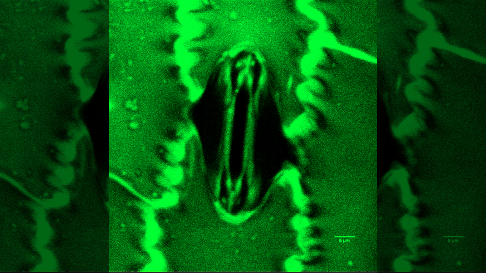
Both electron and light microscopy advanced in the 20th century. Today, labs may use fluorescent tags or polarized filters to view specimens, or they use computers to capture and analyze images that wouldn't be visible to the human eye. There are reflecting microscopes, phase contrast microscopes, confocal microscopes and even ultraviolet microscopes. Modern microscopes can even image a single atom.

 Live Science Plus


