Scientists watch a single electron move during a chemical reaction for first time ever
For the first time, scientists visualized how electrons behave during a chemical reaction, which could help reduce unwanted byproducts in future chemistry.

Get the world’s most fascinating discoveries delivered straight to your inbox.
You are now subscribed
Your newsletter sign-up was successful
Want to add more newsletters?

Delivered Daily
Daily Newsletter
Sign up for the latest discoveries, groundbreaking research and fascinating breakthroughs that impact you and the wider world direct to your inbox.

Once a week
Life's Little Mysteries
Feed your curiosity with an exclusive mystery every week, solved with science and delivered direct to your inbox before it's seen anywhere else.

Once a week
How It Works
Sign up to our free science & technology newsletter for your weekly fix of fascinating articles, quick quizzes, amazing images, and more

Delivered daily
Space.com Newsletter
Breaking space news, the latest updates on rocket launches, skywatching events and more!

Once a month
Watch This Space
Sign up to our monthly entertainment newsletter to keep up with all our coverage of the latest sci-fi and space movies, tv shows, games and books.

Once a week
Night Sky This Week
Discover this week's must-see night sky events, moon phases, and stunning astrophotos. Sign up for our skywatching newsletter and explore the universe with us!
Join the club
Get full access to premium articles, exclusive features and a growing list of member rewards.
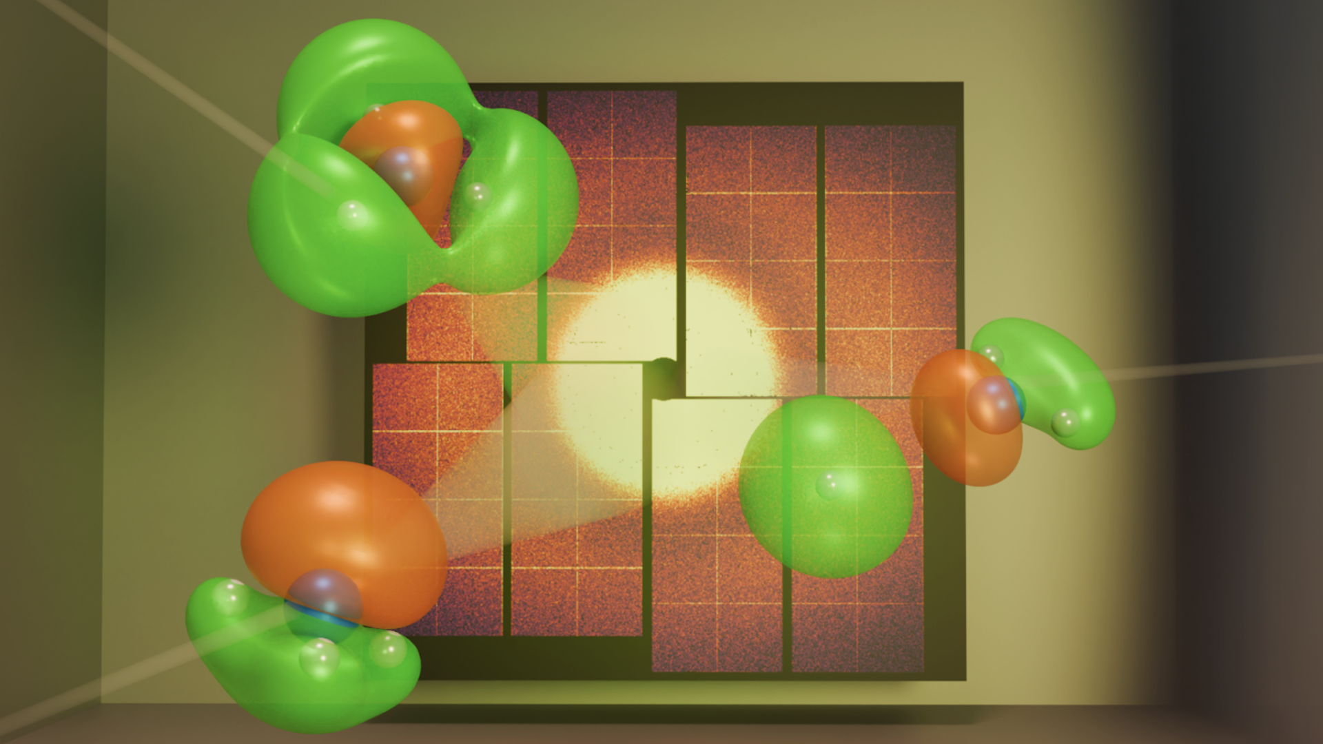
For the first time, scientists have used ultrafast X-ray flashes to take a direct image of a single electron as it moved during a chemical reaction.
In the new study, published Aug. 20 in the journal Physical Review Letters, the researchers accomplished this incredible feat by imaging how a valence electron — an electron in the outer shell of an atom — moved when an ammonia molecule broke apart.
For decades, scientists have used ultrafast X-ray scattering to image atoms and their chemical reactions. The scattering uses supershort bursts of X-rays to freeze tiny, fast-moving molecules in action. X-rays have the perfect wavelength range for capturing details at the atomic scale, which is why they're ideal for imaging molecules.
However, X-rays interact strongly only with core electrons near the atom’s nucleus. Valence electrons — the outermost electrons in an atom and the ones actually responsible for the chemical reactions — were hidden.
"We wanted to take pictures of the actual electrons that are driving that motion," Ian Gabalski, a physics doctoral student and lead author of the study, told Live Science.
If scientists can understand how valence electrons move during chemical reactions, it could help them design better drugs, cleaner chemical processes, and more efficient materials, Gabalski said.
To get started, the team needed to find the right molecule. It turned out to be ammonia.
Get the world’s most fascinating discoveries delivered straight to your inbox.
"Ammonia is kind of special," Gabalski said. "Because it has mostly light atoms, there aren't a lot of core electrons to drown out the signal from the outer ones. So we had a shot at actually seeing that valence electron."

The experiment was conducted at the SLAC National Accelerator Laboratory's Linac Coherent Light Source, a facility that produces intense, short X-ray pulses. First, the team gave the ammonia molecule a tiny jolt of ultraviolet light, which made one of the electrons "jump" to a higher energy level. Electrons in molecules usually stay in low-energy states, and if they are pushed to a higher one, it triggers a chemical reaction. Then, with the X-ray beam, the researchers recorded how the electron's "cloud" shifted as the molecule began to break apart.
Related: The shape of light: Scientists reveal image of an individual photon for 1st time ever
In quantum physics, electrons aren't seen as tiny balls orbiting the nucleus. Instead, they exist as probability clouds, "where higher density means you're more likely to see the electron," Gabalski explained. These clouds are also known as orbitals, and each one has a distinct shape depending on the energy and position of the electron.
To map this electron cloud, the team ran quantum mechanical simulations to calculate the molecule's electronic structure. "So now this program that we use for these kinds of calculations goes and it figures out where the electrons are filling up those orbitals around the molecule," Gabalski said.
The X-rays themselves act like waves, and when they pass through the electron's probability cloud, they scatter in different directions. "But then those X-rays can go and interfere with each other," Gabalski said. By measuring this interference pattern, the team reconstructed an image of the electron's orbital and saw how the electron moved during the reaction.
They compared the results to two theoretical models: one that included valence electron motion, and one that didn't. The data matched the first model, confirming that they had captured the electron's rearrangement in action.
The researchers hope to adapt the system for use in more complex, 3D environments that better mimic real tissues. That would move it closer to applications in regenerative medicine, such as growing or repairing tissue on demand.

Larissa G. Capella is a science writer based in Washington state. She obtained a B.S. in physics and a B.A. in English creative writing in 2024, which enabled her to pursue a career that integrates both disciplines. She reports mainly on environmental, Earth and physical sciences, but is always willing to write about any science that sparks her curiosity. Her work has appeared in Eos, Science News, Space.com, among others.
You must confirm your public display name before commenting
Please logout and then login again, you will then be prompted to enter your display name.
 Live Science Plus


