Wee Wonders: Top 20 Nikon Small World Contest Photos
12th Place, Dr. Dylan Burnette
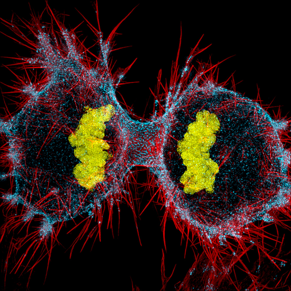
Dr. Dylan Burnette of Vanderbilt University School of Medicine in Nashville, Tennessee, used structured illumination at 9x magnification to offer this image of a human HeLa cell undergoing cell division (cytokinesis) — DNA in yellow, myosin II in blue and actin filaments in red — and received 12th place in Nikon Small World 2016.
13th Place, Walter Piorkowski
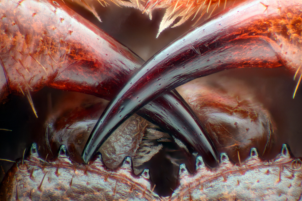
From South Beloit, Illinois, Walter Piorkowski used fiber optic illumination and image stacking at 16x magnification to present the poison fangs of a centipede (Lithobius erythrocephalus) and take home 13th place in the photomicrography contest.
14th Place, Dr. Keunyoung Kim
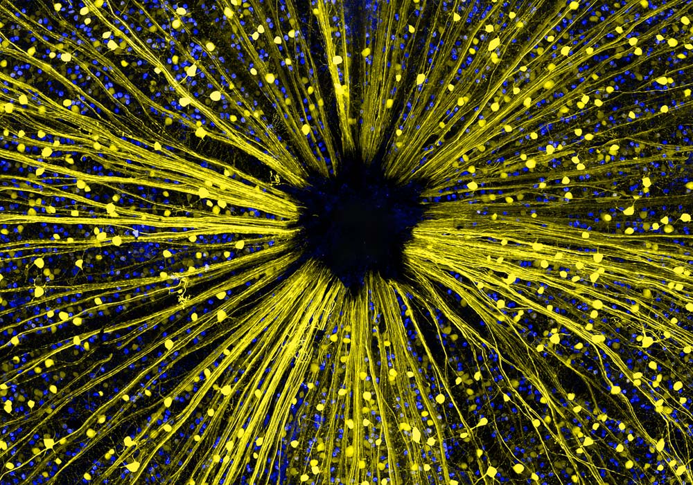
Garnering 14th place in the Nikon Small World 2016 contest, Dr. Keunyoung Kim from the University of California, San Diego, National Center for Microscopy and Imaging Research (NCMIR) in La Jolla, California submitted this image of mouse retinal ganglion cells at 40x magnification using both fluorescence and a confocal lens.
15th Place, Geir Drange
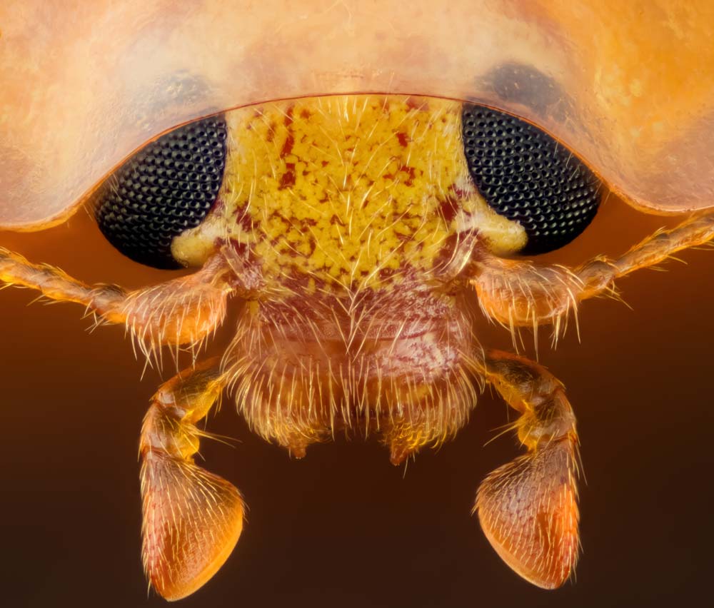
Geir Drange from Asker, Norway, took 15th place in Nikon Small World 2016 with the head section of an orange ladybird (Halyzia sedecimguttata) taken with reflected light and focus stacking at 10x magnification.
16th Place, Stefano Barone
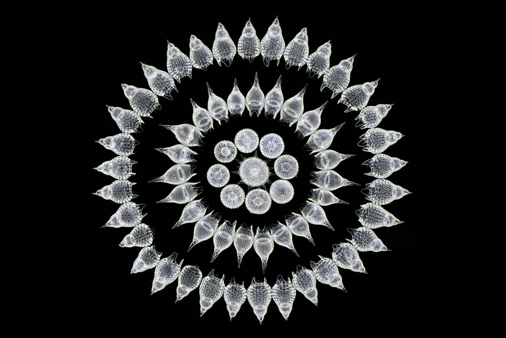
Using darkfield imaging at 100x magnification, Stefano Barone from the Diatom Shop in Palazzo Pignano, Italy, offered this image of 65 fossil Radiolarians (zooplankton) carefully arrange by hand in Victorian style, winning 16th place in the photomicrography contest.
17th Place, Jose Almodovar
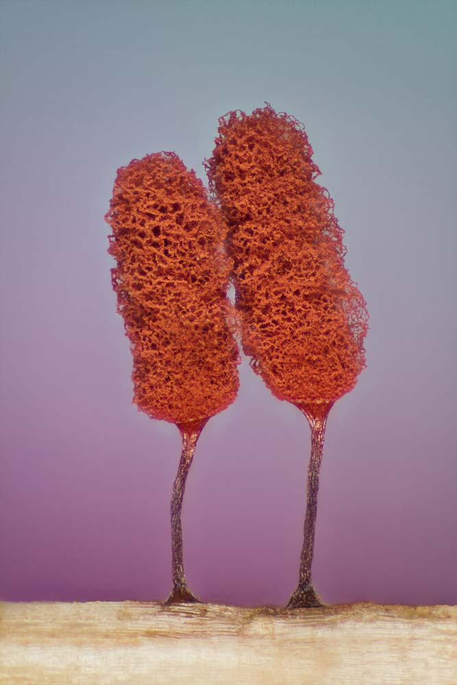
From the Biology Department of the Mayaguez Campus at the University of Puerto Rico in Mayaguez, Puerto Rico, Jose Almodovar took 17th place with this slime mold (Mixomicete) image captured with image stacking and reflected light at 5x magnification.
18th Place, Pia Scanlon
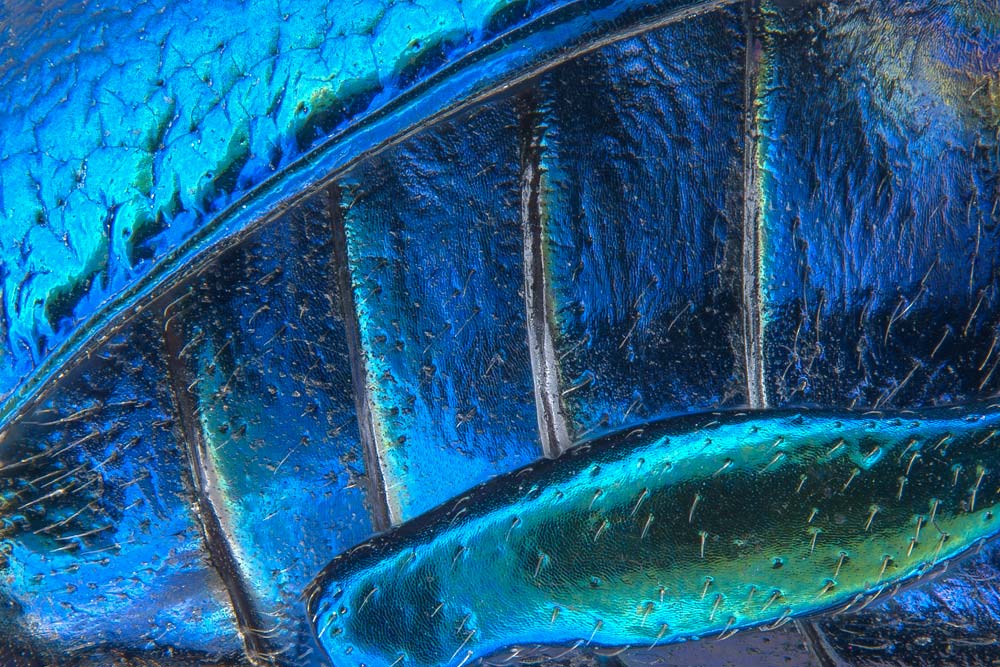
Using stereomicroscopy and image stacking at 40x magnification, Pia Scanlon with the Department of Agriculture and Food, Western Australia, Biosecurity and Regulation - Pest Diagnostics inSouth Perth, Western Australia, took home 18th place in the Nikon Small World 2016 contest with this image of parts of wing-cover (elytron), abdominal segments and the hind leg of a broad-shouldered leaf beetle (Oreina cacaliae).
Get the world’s most fascinating discoveries delivered straight to your inbox.
19th Place, Dr. Gist F. Croft, Lauren Pietilla, Stephanie Tse, Dr. Szilvia Galgoczi, Maria Fenner, Dr. Ali H. Brivanlou
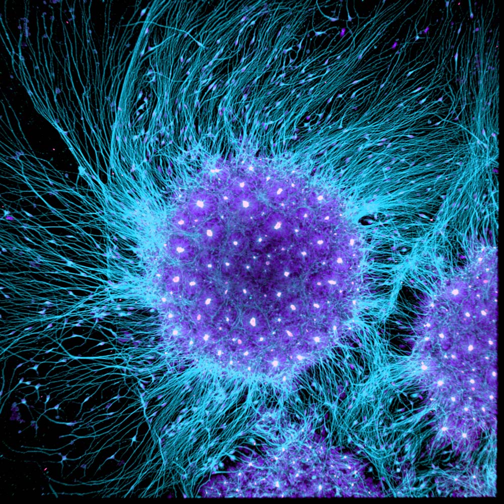
From the Brivanlou Laboratory at Rockefeller University in New York, New York, Dr. Gist F. Croft, Lauren Pietilla, Stephanie Tse, Dr. Szilvia Galgoczi, Maria Fenner and Dr. Ali H. Brivanlou captured 19th place in the Nikon Small World 2016 competition with an image of human neural rosette primordial brain cells, differentiated from embryonic stem cells taken with a confocal lens at 10x magnification.
20th Place, Michael Crutchley
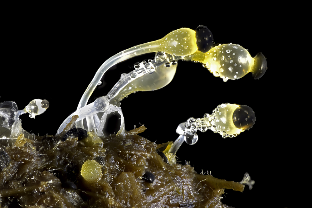
Michael Crutchley of Haverfordwest, Pembrokeshire, UK, offered this interesting image of cow dung using the darkfield method at 30x magnification and captured 20th place in the Nikon Small World 2016 photomicrography competition.


