Live Science Plus


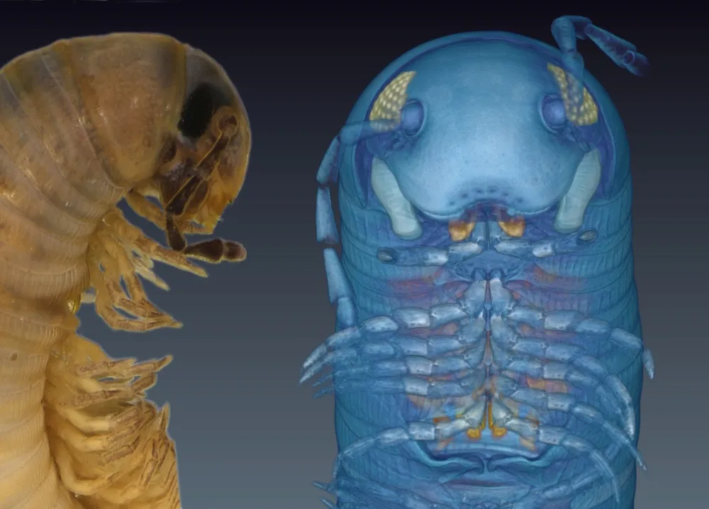
The 3D cyberportrayal of a newfound millipede looks like it hopped from the screen of James Cameron's "Avatar," with a blue body and alien appearance to boot.
The 1.5-inch-long (3.8 centimeters) millipede was discovered in Andalusia in Spain, in 2005. It lives in the dirt underneath stones and leaf litter — a decomposer that "acts as an important component of soil fauna," said study lead researcher Nesrine Akkari, the curator of the Myriapoda Collection at the Museum of Natural History in Vienna.
After preserving the specimen in a jar of ethanol for several years, the researchers decided to introduce it to the scientific world in a novel way. Newfound species are typically photographed, illustrated or put under a microscope, but in this case — for the first time — the researchers used X-ray microtomography (micro-CT) to study it. [See Stunning 3D Images of the Millipede's Avatar]
Article continues belowMicro-CT is essentially the same as a medical computerized tomography (CT) scanner, except that it's more detailed, with a resolution down to a few micrometers, Akkari said. The researchers placed the jar containing the millipede in the machine, which then took X-rays of the specimen from different angles. When the X-rays were compiled, the researchers created a detailed 3D avatar of the millipede, named Ommatoiulus avatar.
"We decided to bring some innovation to the study and show that taxonomy [the classification of species] is not an old-fashioned kind of discipline, but that it can be fun, it can be modern," Akkari told Live Science.
The finding is the first new species to be described with the help of micro-CT imaging, and to be published with its so-called cybertypes for free viewing online.
Avatar advantages
The researchers not only created a 3D avatar of the millipede — which looks like a weird blue teddy bear, but with hundreds of legs — but also managed to keep the specimen intact. If they had analyzed it in the traditional way, they would have needed to slice and dice the arthropod to study its anatomy, including its internal organs.

The technique also allowed the researchers to get an unprecedented look at the millipede's genital appendages, known as the gonopods. Gonopod anatomy varies in millipedes, and it can help scientists identify new species.
"Usually, to examine those genitalia, we would have to make a cut, a dissection, and cut the specimen at the seventh ring, extract those gonopods and cut them in half," Akkari said. "And we could virtually do that without touching the [specimen]."
Instead, the researchers returned the unblemished millipedes to the Natural History Museums of Vienna and Denmark, where they can be used for future research, Akkari said.
Researchers have used micro-CT to look at other specimens, including insects such as bees, the reptilian tuatara and even the charred remains of a 1,500-year-old Hebrew scroll.
"Micro-CT is a great new technique for species descriptions," said Thomas Wesener, the curator for millipedes at the Alexander Koenig Zoological Research Museum in Germany, who was not involved in the study.
"It allows [researchers] to combine the publication of the species name with the online publication of a dataset, including photographs and scans of virtually all morphological characters," Wesener told Live Science in an email. "Researchers will study the dataset for years and compare the scans with those of other millipedes." [Image Gallery: Check Out the Leggiest Millipede]
He noted that other imaging techniques provide even greater detail, including synchrotron micro-CT, where researchers "utilize the beam line of large particle accelerators, such as [Large Hadron Collider at] the CERN in Switzerland, [the Hadron Elektron Ring Anlage at] DESY in Germany or, previously, the Tevatron, close to Chicago."
But these techniques take more time to complete than classical species descriptions do. Wesener estimates that scientists probably need two weeks, as well as technical help, to get raw data, interpret it and create images suitable for publication.
"It might be worth it, because you get so much additional data from the species," he said. "I currently have close to 150 undescribed millipede species in my office, so I probably will not have the time to use micro-CT for every species."
The study was published online today (Aug. 26) in the journal PLOS ONE.
Follow Laura Geggel on Twitter @LauraGeggel. Follow Live Science @livescience, Facebook & Google+. Original article on Live Science.