It's quick and easy to access Live Science Plus, simply enter your email below. We'll send you a confirmation and sign you up for our daily newsletter, keeping you up to date with the latest science news.
A creepy-crawly millipede is the first newfound species to be described with a 3D imaging method called X-ray microtomography (micro-CT). The micro-CT method is similar to that used by a medical CT scanner: It takes X-rays of the specimen from different angles and stacks them on top of one another to create a detailed 3D image. [Read the full story on the millipede imaged with microCT]
Preserved millipede
A preserved male specimen of the millipede (Ommatoiulus avatar). Researchers first discovered the species in the Spanish province of Andalusia, in 2005. Males can measure up to 1.3 inches (33.2 millimeters) in length. Females are slightly longer, reaching up to 1.5 inches (38.2 mm). (Image credit: Akkari N, Enghoff H, Metscher BD (2015) PLoS ONE 10(8): e0135243. doi:10.1371/journal.pone.0135243. Creative Commons.)
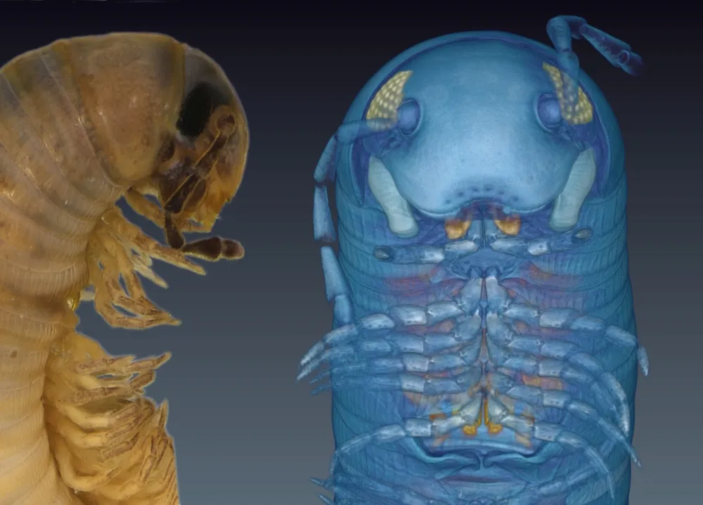
An image of the 3D, micro-CT-generated avatar next to a preserved specimen of Ommatoiulus avatar. The species takes its name from its computer-generated avatar. (Image credit: Licensed Creative Commons by Metscher and Akkari.)
Blue bug
The micro-CT provides a detailed view of the male centipede's gonopods (red and yellow) body parts used in reproduction. Millipedes often have distinct gonopods, allowing researchers to identify newfound species by looking at the gonopods' unique structures. (Image credit: Licensed Creative Commons by Metscher and Akkari.)
Sign up for the Live Science daily newsletter now
Get the world’s most fascinating discoveries delivered straight to your inbox.
Mr. Millipede
Typically, researchers photograph and illustrate new species of animals, sharing the new specimen with the world. Ommatoiulus avatar is the first newfound species of millipede to receive 3D imaging treatment for its scientific debut.
Here is an image of the male avatar. Notice its gonopods (orange), including a pair of modified legs in its mouth region that help transfer sperm packets to the female during mating. (Image credit: Akkari N, Enghoff H, Metscher BD (2015) PLoS ONE 10(8): e0135243. doi:10.1371/journal.pone.0135243. Creative Commons.)
Male gonopod
An image made with a scanning electron microscope of the male millipede's gonopod. (Image credit: Akkari N, Enghoff H, Metscher BD (2015) PLoS ONE 10(8): e0135243. doi:10.1371/journal.pone.0135243. Creative Commons.)
Ms. Millipede
The female avatar's vulvae and eggs are highlighted in this 3D image. (Image credit: Akkari N, Enghoff H, Metscher BD (2015) PLoS ONE 10(8): e0135243. doi:10.1371/journal.pone.0135243. Creative Commons.)
Virtual slice
A virtual section of the cybertype male (A and C), showing its gonopods, musculature and other anatomical features. The cybertype female (B) shows the vulvae, eggs, musculature and other internal structures. (Image credit: Akkari N, Enghoff H, Metscher BD (2015) PLoS ONE 10(8): e0135243. doi:10.1371/journal.pone.0135243. Creative Commons.)
Laura is the managing editor at Live Science. She also runs the archaeology section and the Life's Little Mysteries series. Her work has appeared in The New York Times, Scholastic, Popular Science and Spectrum, a site on autism research. She has won multiple awards from the Society of Professional Journalists and the Washington Newspaper Publishers Association for her reporting at a weekly newspaper near Seattle. Laura holds a bachelor's degree in English literature and psychology from Washington University in St. Louis and a master's degree in science writing from NYU.

 Live Science Plus