
 Live Science Plus


Stunning Photos Reveal Beauty in Medicine

The 14th annual Wellcome Image Awards were presented on March 18, highlighting 20 incredible medical images that ranged from the uterus of a pregnant pony and a cat's tongue to a mouse brain and greenfly eye. Here's a look at the stunning images and why the judges chose them. [Read the full story on the Wellcome Image Awards]
My pregnant pony
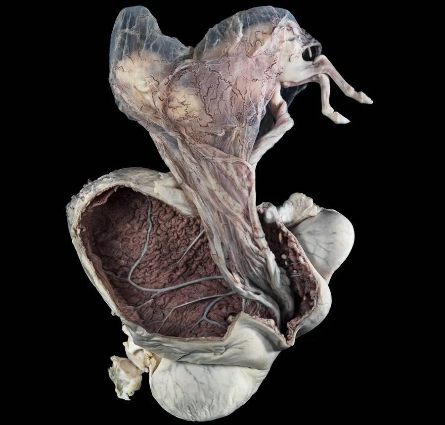
Photograph of a pregnant uterus (womb) from a New Forest pony, approximately five months into the pregnancy. The developing pony (fetus) is outside the uterus but remains attached by its membranes and umbilical cord. The bent back legs of the fetus are sticking out from the membranes (top right-hand side). The uterus has been cut open to reveal its vast blood supply, which is visible on the inner surface. This historical specimen is from a cull animal that happened to be pregnant at the time. It is preserved in formalin in a Perspex container and was photographed in the Anatomy Museum of the Royal Veterinary College in London. (Credit: Michael Frank, Royal Veterinary College / Wellcome Images)

Tim Burton figure?

Micro-computed tomography (micro-CT) scan of the skull and front legs of a tuatara. The tuatara of New Zealand are all that remain of a group of animals that used to share the earth with dinosaurs. Their name comes from the spines along their neck, back and tail; Tuatara is a Maori word meaning ‘spiny back’. Virtual X-ray ‘slices’ of a preserved specimen of this rare reptile were taken and used to create a digital 3D model. The digital model can be virtually cut and rotated without damaging the precious original sample. In this way, the sesamoid bones (tiny bones within tendons in the limbs) can be easily visualised and studied. (Credit: Sophie Regnault, Wellcome Images)
Whimsical grains

Illustration of pollen grains being released from a flower in the Asteraceae family. Asteraceae is one of the largest families of flowering plants and is commonly known as the aster, daisy, sunflower or composite family. Pollen grains contain the male sperm cell and are produced in the anther, one of the male parts of the flower. They are carried to other flowers – primarily by insects, birds and the wind – so flowering plants can reproduce. They look like fine dust and are a common cause of hay fever or seasonal allergies. (Credit: Maurizio De Angelis, Wellcome Images)
Get the world’s most fascinating discoveries delivered straight to your inbox.
Mouse lungs

Confocal micrograph of whole mouse lungs (blue and green). Microparticles that can carry medicines (pink) are being studied to see whether they can deliver these drugs to the lungs. Current anticancer therapies have many toxic side-effects, so researchers hope that these microparticles could one day deliver anticancer medicine in a much simpler, more targeted way – for example, in an inhaler – with fewer side-effects. As microparticles release drug over time, fewer doses may be needed. The right lung in mice is divided into four lobes (right side of image), but the left lung has only one lobe (left side of image). Remnants of the windpipe (trachea) and surrounding tissue are also visible (center). (Credit: Gregory Szeto, Adelaide Tovar and Jeff Wyckoff, Wellcome Images)
Cat tongue

A polarized light micrograph shows a cross-section of part of a cat's tongue. The round protrusions are papillae give the tongue its rough sandpaperlike texture that helps a cat pick up food and clean its fur. This tongue sample came from a vintage slide prepared in the Victorian era. "This striking image looks a bit like bubbling lava, but is in fact the barbed surface of a cat’s tongue. If you’ve ever had a cat lick your hand and wondered why it feels like sandpaper, then this explains it," one of the judges, Fergus Walsh, medical correspondent for the BBC, said in a statement. "Sometimes images that show nature in extreme close-up are both beautiful and illuminating." (Credit: David Linstead, Wellcome Images)
Star Wars character?

Scanning electron microscope composite image of the head of a boll weevil (Anthonomus grandis) found on the front porch of a suburban house. The boll weevil is a beetle that feeds on and lays its eggs in the cotton plant. These agricultural pests have long curved snouts (often half as long as their bodies) and can destroy entire cotton crops. They develop from egg to adult in approximately 20 days and grow on average to 6–8 mm in length. This is one image in a series of work looking at common household pests found inside homes on the outskirts of town. These images of our often-overlooked housemates are in the style of traditional portraits. (Credit: Daniel Kariko, Wellcome Images)
Mouse brain

Confocal micrograph of nerve cells inside a section of adult mouse brain. The brain has been sliced (like a loaf of bread), and one of those pieces is seen here. After being sliced, it was chemically treated to make the tissue transparent so that structures deep inside could be more easily seen. A subset of nerve cells tagged with a visual marker (green fluorescent protein) were visualised at different depths through the piece of tissue, which is 0.75 mm thick. The markers are colour coded from red (nearest) to orange, yellow, purple, blue and green as you look into the image. (Credit: Luis de la Torre-Ubieta, Wellcome Images)
Teensy wasp

Light micrograph of a tiny parasitoid wasp (Wallaceaphytis kikiae) viewed from above. Parasitoid wasps lay their eggs inside other insects. After hatching, the larvae feed on their host, eating it alive from the inside out. This is a new genus of parasitoid wasp recently discovered in the rainforests of Borneo, where a single female wasp was found mixed in with thousands of other insects. It measures only 0.75 mm in length and has unusual antennae, legs and wings. It’s named after Alfred Russel Wallace, who coauthored the first ever publication on evolution by natural selection with Charles Darwin and who himself identified new insects while in Borneo in the mid-19th century. (Credit: Andrew Polaszek, Wellcome Images)
Brain cell

Scanning electron micrograph of a single brain cell (colored green and pink). A rectangular cut has been made in the cell to watch how tiny, nanometer-sized particles (coloured red and brown) interact with its surface. These tiny particles are called carbon nanotubes and are nano-sized cylinders made of carbon atoms. They are being researched for their ability to act as carriers to deliver drugs or genes to cells – for example, anticancer medicines to a tumour. This is particularly important in the brain because many medicines cannot easily cross the blood–brain barrier, a protective layer of cells that regulates entry of molecules to the brain. (Credit: Khuloud T Al-Jamal, Serene Tay and Michael Cicirko , Wellcome Images)
Nervous system

Reminiscent of a Jackson Pollock painting, this image shows part of the central nervous system in a fruit fly (Drosophila melanogaster). Transmission electron micrographs were used to create a digital colour-coded map of the area. An organism’s nervous system controls everything it does, from breathing and moving to thinking and feeling. Instructions to perform these tasks are carried by cells called neurones. A neurone able to sense vibrations (yellow) is surrounded here by lots of other neurones, each depicted as a single line. Messages enter (blue circles) and exit (red circles) neurones at points of contact called synapses. Other features of interest (orange circles), such as mitochondria, are also marked. (Credit: Albert Cardona, Wellcome Images)
Goat stomach chamber

This image shows the reticulum of a goat. The reticulum is one of the chambers of the ruminant (cattle, goats and sheep) stomach and is characterised by the honeycomb pattern which lines the inside. Bacteria are present here which causes fermentation of the food material. The oesophagus can be seen entering the reticulum at the top of the picture. The opening in the centre leads to the omasum, another chamber of the stomach. A small part of the rumen, another chamber, is attached to the reticulum on the right. The lining changes here to small papillae, and resembles carpet. A goat's stomach has four chambers: the rumen, the reticulum, the omasum and the abomasum. (Credit: Michael Frank, Royal Veterinary College, Wellcome Images)
Attack cells

3D structured illumination micrograph (3D-SIM) of a natural killer (NK) cell (left) attacking a susceptible target cell (less bright, slightly rounder cell on the right). NK cells are key players of the innate mammalian immune system and are capable of recognising and destroying infected cells. NK cells produce toxic substances (cytotoxic) which when delivered to a susceptible target cell causes it to self-destruct (apoptosis or programmed cell death). These cytotoxic substances (pro-apoptotic factors) are stored in specialised compartments (cytotoxic granules) inside the NK cell so that NK cells are always pre-armed and ready to kill. In this image, the NK cell has docked onto the target cell creating an area of contact between the two cells. This tightly sealed, highly organised interface is called the immune synapse. It is here at the immune synapse that focused, controlled release of cytotoxic granules occurs. Filamentous actin (an important component of the cellular cytoskeleton) is shown in grey and cytotoxic granules in red (perforin staining). Scale bar (white; lower left) is 5 micrometres. 3D-SIM is one type of super-resolution microscopy. (Credit: N. Dieckmann and N. Lawrence, Universtiy of Cambridge, Wellcome Images)
3D printed lungs and ribcage

Human lungs inside their ribcage 3D printed in white SLS nylon. In this image, the lungs and ribcage are viewed from the back with the vertebrae of the spine clearly visible in the centre. The lungs and ribcage belong to Caroline who was diagnosed with Hodgkin lymphoma cancer. The 2D data contained in her CT scans were converted into 3D renders by the artist who was then able to export them to a printable format. Original size of artwork is 14h x 13.5w x 9.5d cm. Wellcome Trust photography by Ben Gilbert. (Credit: Dave Farnham, Wellcome Images)
Rat brain

Scanning electron micrograph of the dendritic tree of a purkinje neurone in the cerebellar cortex of a rat brain. Dendritic trees are the tiny receiving elements of neurones and receive the vast majority of input information coming into the neurone. They have an elaborate branching structure which helps to define the 'personality' of individual neurones. Dendrites are not passive receivers, but instead actively transform the signals they receive, which dramatically enhances the computational power of single neurones. The cerebellar cortex forms part of the cerebellum, the region of the brain which plays a role in controlling accuracy and coordination of movement. This purkinje cell was filled with a visual marker using the patch-clamp technique. It was then imaged using a focused ion beam scanning electron microscope which allows neurones and neural circuits to be reconstructed in high resolution. This image is 110 micrometres wide and 141 micrometres high. (Credit: Prof. M. Hausser, Sarah Rieubland and Arnd Roth, UCL, Wellcome Images)
Greenfly eye

Scanning electron micrograph of a greenfly eye. Greenflies (aphid) have a pair of compound eyes for accurate perception of movement. The small protrusion coming from the side of the eye is called an ocular tubercle which is made up of three lenses (triommatidium). Aphids are insects, many species of which are agricultural pests that feed on plant sap. Horizontal width of image is 280 micrometres. (Credit: Kevin Mackenzie, University of Aberdeen, Wellcome Images)
Healthy human brain

Sagittal view of a healthy adult, living human brain, virtually sliced down a vertical axis dividing it into left and right halves. The front of the head is facing towards the left side of the image. Information on neural tracts or pathways of white matter in the brain were collected by a type of magnetic resonance imaging (MRI) which tracks the movement of water molecules within tissue, similar to diffusion tensor imaging (DTI). This was used to reconstruct a digital model in the style of a Joseph Jules Dejerine anatomical drawing. This method of tractography is being used to map networks inside the brain to use as a tool for teaching neuroanatomy and to build atlases of the human brain. Field of view is 256 x 140 mm. (Credit: Dr. Flavio Dell'Acqua, Wellcome Images)
Kyphosis

Kyphosis is a condition causing curvature of the dorsal spine or 'rounding' of the back and shoulders most commonly seen in elderly women. It can be congenital or it can be caused by a number of degenerative and endocrine diseases. It can also occur as a result of trauma. A patient with Kyphosis (also known as dowager's hump) may experience intense back pain and have difficulty breathing. If deemed necessary surgical intervention can be used. This image won the 2014 Wellcome Photographic Award at the 2014 Institute of Medical Illustrators Conference. (Credit: Mark Bartley, Cambridge University Hospitals NHS Foundation Trust, Wellcome Images)
Pediatric distractions

An interactive multi-sensory unit designed to provide a distraction for anxious children undergoing painful hospital procedures. The unit is approximately 5 ft tall and includes a bubble tube, fibre optic lights, mirrors, a solar projector and the capability of producing sound. (Credit: Geraldine Thompson, CMFT, Wellcome Images)
Old anatomy model

This anatomical model was about to be discarded as rubbish when the photographer decided to rescue it and take one last photograph to honour the service it provided to medical students in Trinity College Dublin. The photographer back-lit the model to provide a certain effect and therefore the model looks to be sleeping or taking a rest in a chair after a weary day. (Credit: Anthony Edwards, Wellcome Images)
