Live Science Plus


You are now subscribed
Your newsletter sign-up was successful
Want to add more newsletters?

Delivered Daily
Daily Newsletter
Sign up for the latest discoveries, groundbreaking research and fascinating breakthroughs that impact you and the wider world direct to your inbox.

Once a week
Life's Little Mysteries
Feed your curiosity with an exclusive mystery every week, solved with science and delivered direct to your inbox before it's seen anywhere else.

Once a week
How It Works
Sign up to our free science & technology newsletter for your weekly fix of fascinating articles, quick quizzes, amazing images, and more

Delivered daily
Space.com Newsletter
Breaking space news, the latest updates on rocket launches, skywatching events and more!

Once a month
Watch This Space
Sign up to our monthly entertainment newsletter to keep up with all our coverage of the latest sci-fi and space movies, tv shows, games and books.

Once a week
Night Sky This Week
Discover this week's must-see night sky events, moon phases, and stunning astrophotos. Sign up for our skywatching newsletter and explore the universe with us!
Join the club
Get full access to premium articles, exclusive features and a growing list of member rewards.
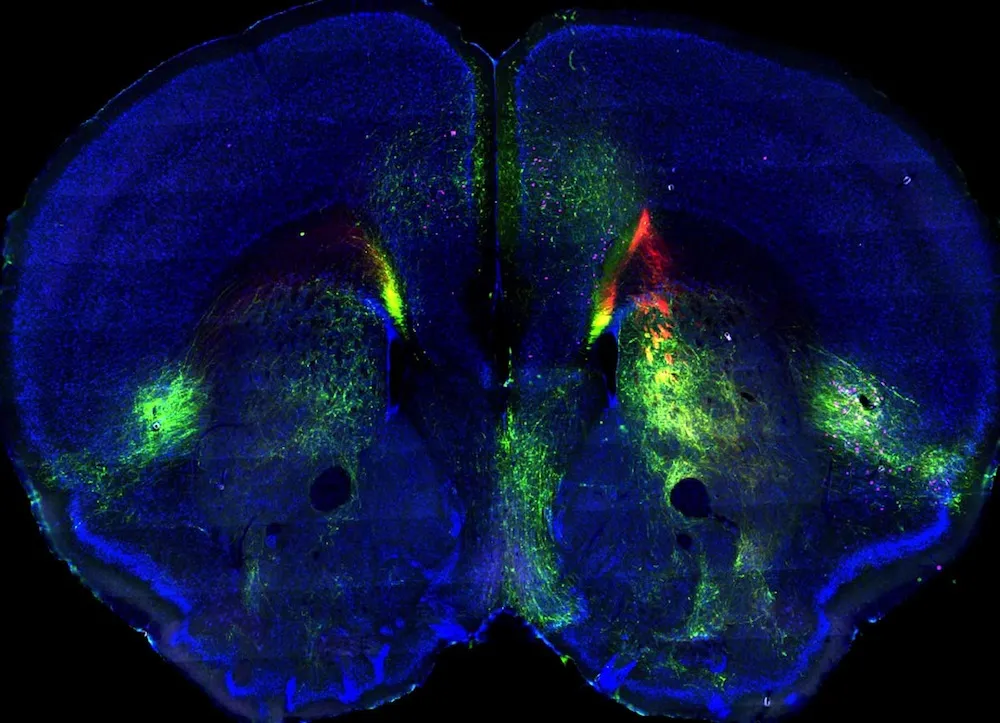
Glowing new images of the mouse brain represent the most comprehensive mapping yet of the mammalian cortex.
Using fluorescent injections, researchers tracked the connections between regions of the mouse cortex, the outermost, wrinkled layer of the brain.
The project is important because the mouse brain is structured basically like other mammal brains — including humans', said study leader Hong-Wei Dong, a neuroscientist at the University of Southern California. Understanding how healthy brain structures chat back and forth should help researchers figure out how to fix problems when something goes wrong. [Inside the Brain: Images Through Time]
"Our ultimate goal is really to understand what is happens in all of these neurological or neuropsychiatric disorders, like autism or schizophrenia," Dong told Live Science.
Mapping the brain
Neuroscientists have increasingly focused on how brain regions connect in order to explain behavior, cognition and even disease. In previous studies, researchers have built brain maps for rats and primates by pulling together thousands of individual research studies on small portions of the brain. What Dong and his colleagues have done, however, is to collect a vast amount of data themselves. Their maps are based on only male mice of the same age, and allow for far more detail than earlier approaches.
"Our strength here is we have a collection of a huge amount of data, and we can then analyze them systematically," Dong said.

The researchers injected fluorescent molecules in two locations in each of 300 mouse brains. These tracers travelled along the neuronal connections, showing which networks of brain cells were sending out signals to where, and which networks were answering back.
"The tracer in that injection site will tell you what areas that structure projects to and what areas project back to that structure," said Houri Hintiryan, a USC neuroscientist who was the co-lead researcher on the study with Dong. Capturing both incoming and outgoing messages is useful scientifically and allows researchers to use fewer mice, Hintiryan told Live Science.
Logical organization
The tracers revealed a methodical organization in the cortex.
"The brain is not randomly wired together," Hintiryan said. "There is a specific logic to its organization."

The mouse cortex is organized into four somatic sensorimotor subnetworks, two medial subnetworks and two lateral subnetworks, Dong said. The somatic sensorimotor subnetworks each have their own functions, as determined from their connectivity: One controls facial movement, one the upper limb, one the lower limb and one the whiskers.
The medial subnetworks, so named because they sit along the midline of the brain, seem to integrate external information, such as information from the eyes and ears. One of the lateral networks handles sensory information from the body itself, including sensations such as hunger, cold and pain. The final lateral network seems to be a very complex center where information from all over the cortex converges.
The researchers make their maps available freely online at www.mouseconnectome.org, and they plan to carry out similar work on the rest of the brain. Dealing with the massive amount of data generated by even pipsqueak mouse brains is a challenge, so they also hope to develop better tools to cope.
The project could inform the large-scale BRAIN Initiative, launched by President Obama in April 2013 with the goal of understanding how human brain networks function. After studying the brain connections of normal mice, Hintiryan said, researchers can then compare the healthy connectivity within the brains of mice with the rodent versions of Alzheimer's, Huntington's and other neurological disorders.
The findings appear online today (Feb. 27) in the journal Cell.
Follow Stephanie Pappas on Twitter and Google+. Follow us @livescience, Facebook & Google+. Original article on Live Science.