Live Science Plus


A major pathway of the human brain involved in visual perception, attention and movement — and overlooked by many researchers for more than a century — is finally getting its moment in the sun.
In 2012, researchers made note of a pathway in a region of the brain associated with reading, but "we couldn't find it in any atlas," said Jason Yeatman, a research scientist at the University of Washington's Institute for Learning and Brain Sciences. "We'd thought we had discovered a new pathway that no one else had noticed before."
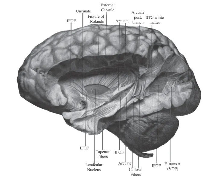
A quick investigation showed that the pathway, known as the vertical occipital fasciculus (VOF), was not actually unknown. Famed neuroscientist Carl Wernicke discovered the pathway in 1881, during the dissection of a monkey brain that was most likely a macaque. [10 Things You Didn't Know About the Brain]
Article continues belowBut besides Wernicke's discovery, and a few other mentions throughout the years, the VOF is largely absent from studies of the human brain. This made Yeatman and his colleagues wonder, "How did a whole piece of brain anatomy get forgotten?" he said.
The researchers immersed themselves in century-old brain atlases and studies, trying to decipher when and why the VOF went missing from mainstream scientific literature. They also scanned the brains of 37 individuals, and found an algorithm that can help present-day researchers pinpoint the elusive pathway.
The study provides a comprehensive look at the VOF's history, said Dr. Jeremy Schmahmann, a professor of neurology at Massachusetts General Hospital and Harvard Medical School, who was not involved in the new research. Schmahmann co-wrote the book "Fiber Pathways of the Brain" (Oxford University Press, 2006), which describes how the VOF is structured in the brain of a monkey and a human.
The new study confirms the VOF's location in the human brain "and then presents a coherent discussion about how it could be relevant," said Schmahmann, who is also a director of the Laboratory for Neuroanatomy and Cerebellar Neurobiology at Massachusetts General Hospital.
Teacher-student dispute?
The VOF may have been the victim of a disagreement between Wernicke and his famous teacher, Theodor Meynert, a German-Austrian neuroanatomist. Meynert directed the psychiatric clinic at the University of Vienna, and also taught Sigmund Freud and the famed Russian neuropsychiatrist Sergei Korsakoff.
Wernicke is known for his 1874 discovery of Wernicke's area, a region of the brain essential for understanding written and spoken language. After his breakthrough, Wernicke studied in Meynert's lab for about six months in the late 1870s and early 1880s.

But although Wernicke also discovered the VOF, Meynert did not include it in any of his studies. It's possible that Meynert ignored the pathway because it broke one of his tenets about brain organization, Yeatman told Live Science.
"Meynert had proposed the original theory of the organization of these pathways," Yeatman said. "He proposed that, as a rule, they all go anterior-posterior, or basically from front to back, longitudinally across the brain."
The VOF, in contrast, goes up and down. "Wernicke's discovery contradicted this majorly accepted principle of brain organization," Yeatman said.
Other neuroanatomists found the VOF in the human brain, but the pathway sits largely unlabeled in brain atlases throughout history, Yeatman said. [3D Images: Exploring the Human Brain]
Yet maybe Meynert didn't mean any harm, Schmahmann said. Meynert did not focus on fiber pathways in the occipital lobe, including, but not limited to the VOF. "Meynert's apparent non-discussion of these fiber systems may simply have reflected his interest and focus," Schmahmann said.
Moreover, the VOF's also went by many names, which may have pushed it into further obscurity. Atlases give it different labels, including "Wernicke's perpendicular fasciculus," "perpendicular occipital fasciculus of Wernicke" and "stratum profundum convexitatis."
Varying dissection techniques in the late 1800s and early 1900s also made the VOF hard to pinpoint.
"You're slicing with a knife and trying to look for structure. It's very easy to miss something if you slice it a different way," Yeatman said.
Pathway reintroduction
To remedy the confusion, Yeatman and his colleagues wrote an algorithm to help researchers find and identify the VOF. They used an MRI technique called diffusion-weighted imaging, which measures the size and direction of the brain's different pathways.

After imaging the brains of 37 people, the researchers found that the VOF starts in the occipital lobe, a part of the brain that processes visual information. It then spreads out like a sheet, connecting different brain regions: those that help people perceive visual categories, such as words and faces, and those involved with eye movements, attention and motion perception, the researchers said. The pathway could therefore help explain how the brain connects the two types of visual perception, Schmahmann said.
"There has to be some way for that dichotomy to merge," he said, "and the Wernicke fascicle is one way for the 'where' and the 'what' streams in the visual modality to become a unified whole."
Interestingly, two case studies from the 1970s found that people with damage to the VOF lost their ability to read because they could no longer recognize words. Moreover, the VOF has different myelination, a coating on nerve cells that helps information move faster.
"We don't know what it means yet, but [the myelination differences are] very consistent across every subject," Yeatman said. "It opens up some new hypotheses, new directions to study: Why is this structure so different than the other neighboring pathways?"
The study, published today (Nov. 17) in the journal Proceedings of the National Academy of Sciences, may encourage researchers to include the VOF in future brain atlases, Yeatman said.
Follow Laura Geggel on Twitter @LauraGeggel. Follow Live Science @livescience, Facebook & Google+. Original article on Live Science.