Scientists discover never-before-seen part of human cells — and it looks like a snowman wearing a scarf
Scientists say they captured 3D images of a new organelle they're calling a "hemifusome," which may be a recycling center in human cells.

Get the world’s most fascinating discoveries delivered straight to your inbox.
You are now subscribed
Your newsletter sign-up was successful
Want to add more newsletters?

Delivered Daily
Daily Newsletter
Sign up for the latest discoveries, groundbreaking research and fascinating breakthroughs that impact you and the wider world direct to your inbox.

Once a week
Life's Little Mysteries
Feed your curiosity with an exclusive mystery every week, solved with science and delivered direct to your inbox before it's seen anywhere else.

Once a week
How It Works
Sign up to our free science & technology newsletter for your weekly fix of fascinating articles, quick quizzes, amazing images, and more

Delivered daily
Space.com Newsletter
Breaking space news, the latest updates on rocket launches, skywatching events and more!

Once a month
Watch This Space
Sign up to our monthly entertainment newsletter to keep up with all our coverage of the latest sci-fi and space movies, tv shows, games and books.

Once a week
Night Sky This Week
Discover this week's must-see night sky events, moon phases, and stunning astrophotos. Sign up for our skywatching newsletter and explore the universe with us!
Join the club
Get full access to premium articles, exclusive features and a growing list of member rewards.
A new organelle has been discovered in human cells — and scientists call it a "hemifusome."
Like the full-size organs in our bodies, the organelles within cells are specialized structures that carry out specific functions. While observing filaments that maintain the shape of cells, Seham Ebrahim, an assistant professor at the University of Virginia, and her team noticed a new structure that was consistently appearing in the 3D images they were making.
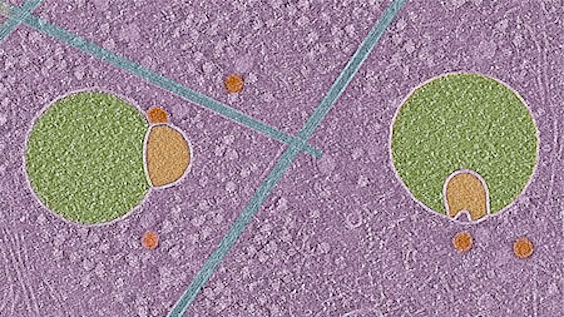
What at first seemed like an artifact in the images turned out to be a new organelle that may be involved in sorting, recycling and discarding proteins within human cells. Ebrahim likened the hemifusome's shape to that of a snowman wearing a scarf; picture a small head attached to a larger body, with a thin border separating the two ends.
The organelle is around 100 nanometers in diameter, less than half the size of even a small mitochondrion, the famous powerhouse of the cell.
The scientists were able to observe the hemifusomes because they used a method called cryo-electron tomography (cryo-ET) to generate their images. This technique involved rapidly freezing cells from four lab-raised cell lines to preserve as much of their structures as possible, making it possible to create clear, 3D images.
"It's like a snapshot in time without any kind of chemical or any kind of stain," Ebrahim told Live Science. Using this imaging technique, they could look inside cells in a "very native state," as if they were glass balls, she said.
Related: Meet the 'frodosome,' a brand new organelle
Get the world’s most fascinating discoveries delivered straight to your inbox.
In their paper, published in the journal Nature Communications in May, the researchers wrote that the harsh processing steps that other imaging techniques subject cells to likely prevented hemifusomes from being observed earlier. In addition, with other techniques that use imaging to study the traffic unfolding in a living cell, the organelle was probably too small to be seen, appearing at most as a blur, Ebrahim added.
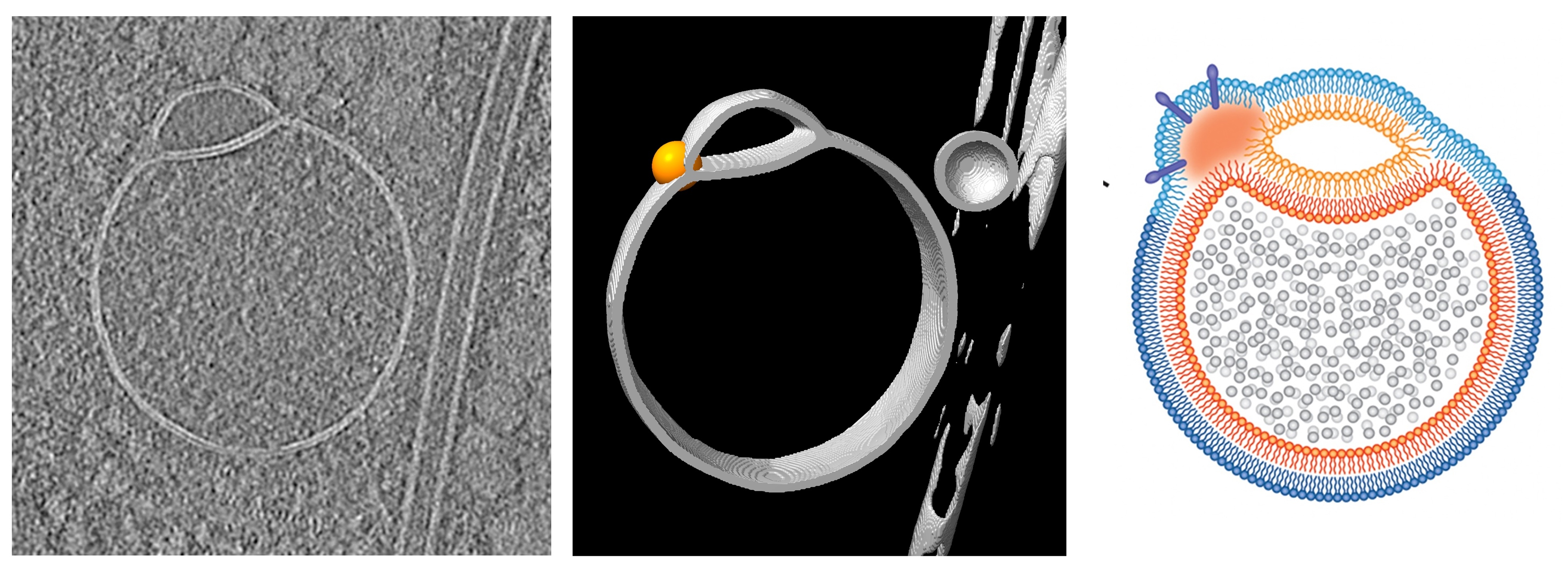
Ebrahim and her colleagues were looking at a configuration of vesicles they had never observed before. Vesicles are balloon-like structures used to transport things like proteins and hormones within and between cells. The new study revealed two vesicles fused together with a two-layer barrier of fat between them.
"Even from a biophysics perspective, it's a breakthrough," Ebrahim said, "because biophysically, people have always predicted, or theorized, that vesicles can exist in this hemifused state … but this was the first time that it has actually been seen in a living cell." This observation inspired the name hemifusome, since hemifusion refers to the partial merger of two bilayers.

Ebrahim argues that hemifusomes can be classified as organelles because they are self-contained functional units within a cell, as opposed to "fleeting" structures that temporarily appear as membranes form and split. In the paper, she wrote that it's unlikely hemifusomes are artifacts of cryo-ET.
Ebrahim's findings "suggest that the hemifusomes they visualize are genuine cellular intermediates, not freezing-induced distortions," said Yi-Wei Chang, an assistant professor of biochemistry and biophysics at the University of Pennsylvania Perelman School of Medicine who was not involved in the work.
Once the role and function of hemifusomes have been confirmed through further studies, they may be recognized as their own class of intermediate structures that fulfill part of fusion processes in mammalian cells, Chang told Live Science in an email.
With their current work, the researchers can confirm that the hemifusome exists, but they have yet to determine the organelle's exact role, its life cycle or its composition. Ebrahim hypothesizes that hemifusomes are precursors to certain types of vesicles. She believes hemifusomes may play a crucial role in the recycling or disposal of cellular membranes, which is important for preventing the buildup of stuff in cells that could gum up their operations if allowed to accumulate.
The researchers wrote that understanding more about how hemifusomes work could also unlock new insight into how diseases such as Alzheimer's manifest. Alzheimer's disease is linked to the improper clearance of abnormal protein plaques in the brain, which leads to buildup over time.
"Without cryo-electron tomography, we would have missed this discovery," Ebrahim said, adding that "there's probably a whole world out there that we still have to find."

Christoph Schwaiger is a freelance journalist, mainly covering health, technology, and current affairs. His stories have been published by Live Science, New Scientist, BioSpace, and the Global Investigative Journalism Network, among other outlets. Christoph has appeared on LBC and Times Radio. Additionally, he previously served as a National President for Junior Chamber International (JCI), a global leadership organization, and graduated cum laude from the University of Groningen in the Netherlands with an MA in journalism.
You must confirm your public display name before commenting
Please logout and then login again, you will then be prompted to enter your display name.
 Live Science Plus


