Scientists stumble upon a new part of a cell in one of the most studied animals on Earth
Scientists found a previously unrecognized organelle in fruit flies, a thoroughly studied organism.

A phosphate-regulating organelle has been discovered in animals for the first time. Until now, only bacteria, yeast and plants have been known to have comparable features.
Despite scientists studying fruit flies (Drosophila melanogaster) for more than a century, the newfound organelle has only just been discovered in the insect. Now researchers are taking a retrospective look at older data in search of these elusive cell parts.
Organelles are microscopic structures inside of cells that perform specialized functions, a variety of which involve phosphate — an essential nutrient for metabolism, storing chemical energy and synthesizing DNA.
"In terms of intracellular regulation of phosphate [in animals], very little is known," said Charles (Chiwei) Xu, a geneticist formerly at Harvard Medical School and first author of a new report describing the discovery of the fly organelle. The report, published May 3 in the journal Nature, notes that the organelle, found in the fruit fly gut, sequesters phosphate from food and regulates its availability in the cell.
Related: Meet the 'frodosome,' a brand new organelle
The fruit fly is one of the most thoroughly researched model organisms, or non-human species used to study fundamental biology.
"It's quite amazing that, in model organisms, we are still discovering things every day that nobody suspected before," Laurent Seroude, a geneticist at Queen's University in Canada told Live Science.
Get the world’s most fascinating discoveries delivered straight to your inbox.
Seroude, who was not involved with the work, noted that discoveries made in model organisms often apply to other species, so it's possible that other animals carry the new organelle. But for now, this is speculation.
Xu and his colleagues' work started out studying how phosphate absorption during digestion affected tissue renewal in fruit flies' guts. When they fed flies meals low in phosphate or gave flies a drug that inhibited phosphate absorption, the researchers noticed something counterintuitive: Despite having little phosphate, the cells lining the fruit fly's gut multiplied rapidly. The cells also multiplied furiously when the team suppressed a protein known to govern phosphate transport inside cells, called PXo.
To explore PXo's role further, the team fused it to a fluorescent protein and peered through a fluorescence microscope to find that it was located on oval-shaped structures in the cell. This was when the team made their chance discovery: They used various stains for known organelles to try to identify these oval structures, but they found that no stains worked and realized they'd stumbled upon a new organelle.
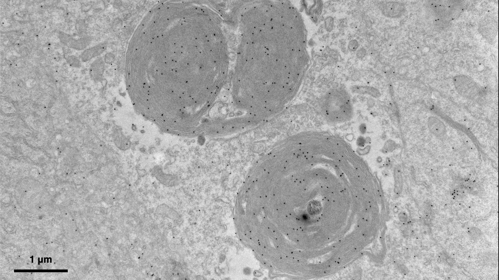
They named the newfound organelles "PXo bodies" and used electron microscopy to study their architecture, revealing membrane whorls, or spirals. These whorls were studded with PXo proteins that transport phosphate from the cytoplasm — the fluid surrounding organelles — into the PXo bodies for storage, thereby regulating the supply of phosphate available for cellular functions.
"The gut is a prominent tissue for nutrient absorption," which may explain why PXo bodies were mainly found there, where they could control phosphate supply to the rest of the body, said Xu.
Seroude said the results were rigorous because the team "systematically used different approaches to show the same thing," namely, that the PXo protein was inextricably linked to the newly discovered organelles. However, he was not convinced that cell division speeds up when an essential nutrient for growth is limited and argued that this hypothesis needs more testing. Xu argued, however, that cell renewal may help boost phosphate absorption when there's a deficit.
"I wouldn't say that we found this organelle from out of nowhere," Xu remarked, but new research techniques allowed his team to characterize the membrane whorls that had previously been overlooked.
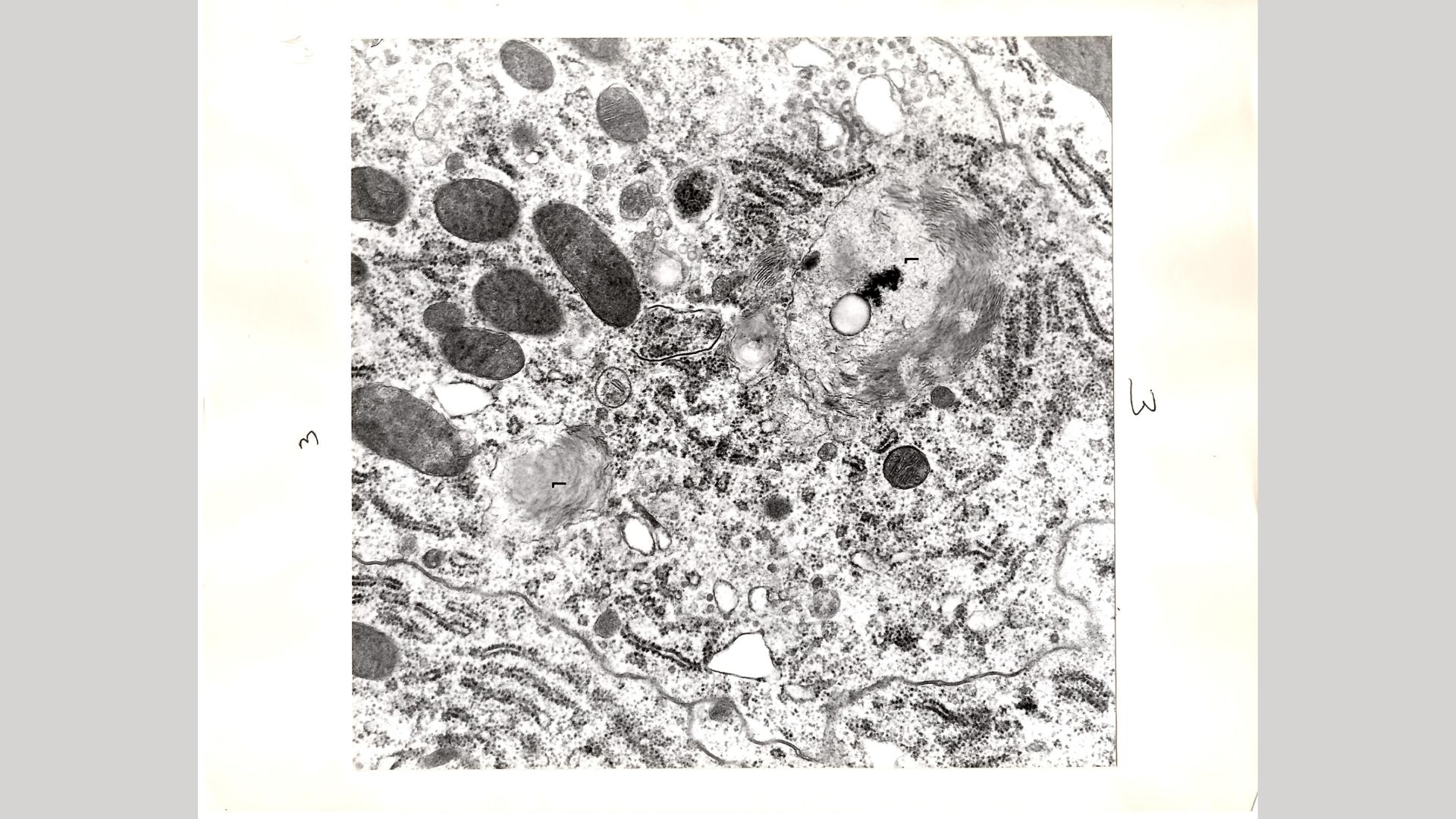
Since the Nature report, researchers have reached out to Xu to share images of organelles that resemble PXo bodies.

One such researcher was Leslie Gartner, a retired cell biologist who emailed Xu about an old study of his: "My study occurred more than 50 years ago, so it was dead and buried beneath the thousands of Drosophila research articles and it would have been almost miraculous had you been able to discover it." Gartner added that he hasn't seen anything like these structures in the fly gut until now.
Xu and his colleagues have identified several proteins that interact with PXo and they plan to decipher what roles they play in regulating phosphate transport into the newly named organelle.

Kamal Nahas is a freelance contributor based in Oxford, U.K. His work has appeared in New Scientist, Science and The Scientist, among other outlets, and he mainly covers research on evolution, health and technology. He holds a PhD in pathology from the University of Cambridge and a master's degree in immunology from the University of Oxford. He currently works as a microscopist at the Diamond Light Source, the U.K.'s synchrotron. When he's not writing, you can find him hunting for fossils on the Jurassic Coast.
 Live Science Plus


