A new imaging technique is giving scientists a rare and precious glimpse into the three-dimensional structure of tiny unborn creatures that were frozen in time more than 500 million years ago.
Called synchrotron-radiation X-ray tomography, the technology was used to glimpse the hidden interiors of embryos from two ancient worm-like species, called Pseudooides and Markuelia. Both fossils are less than a millimeter across and were found in China and Siberia.
The embryos are the oldest full embryonic body fossils of any complex animal ever discovered and among the earliest embryos known. They are predated only by a 580-million-year-old embryo-like structure belonging to an organism thought to be part of the lineage leading to animals.
Latest Videos From
Sifting for fossils
Before the fossils could be imaged, they had to be picked out from about 12 metric tons of rock, a process that took six years, said study team member Phil Donoghue of the University of Bristol in England.
"We picked through every single grain to determine whether it was sand or embryos," Donoghue told LiveScience.
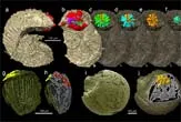
The new technique revealed various developmental stages of the fossilized embryos, from the splitting of cells to pre-hatching. Researchers also got a detailed look at the internal anatomy of the mouth and anus of the unborn Markuelia and found a unique pattern for making embryonic worm segments in Pseudooides not seen in any living animal today.
The images, detailed in the Aug. 9 issue of the journal Nature, also revealed characteristics within one of the embryos similar to those of modern arthropods, the phylum that includes insects, crustaceans, spiders and millipedes. The finding implies arthropod evolution began a few million years earlier than previously thought, the researchers say.
A new tool
Because of their tiny size and fragility, embryos are among the rarest types of fossils found.
"They are just gelatinous balls of cells that rot away within hours," Donoghue said. "But these fossils are the most precious of all because they contain information about the evolutionary changes that have occurred in embryos over the past 500 million years."
The oxygen-poor sediments where the embryos were found likely contributed to their preservation, safeguarding them until minerals could replace their delicate structures, Donoghue said.
The new technology is the latest to allow scientists to study fossils in detail without destroying them. In another recent study, researchers used laser technology to take 3D snapshots of fossils trapped in rock without having to crack the rocks open.
Previously, scientists had to settle for studying parts of fossils that were exposed or they had to slice the specimens, giving only two-dimensional views.

 Live Science Plus