
 Live Science Plus


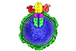
Researchers have developed a three-dimensional model that catches a virus in the act of infecting a host cell.
The model is one of the most detailed images of the tiny infectious agents ever produced. It shows a virus called Epsilon 15 poised to inject a long coil of DNA down a cylinder into a Salmonella bacterium.

The researchers photographed about 15,000 frozen viruses and fed the images into a computer. The images were then used to construct a 3D-model based on common features.
A team of researchers from Northwestern University also recently created a 3D virus model using another technique, called X-ray crystallography.
The latest study, led by Wen Jiang and Wah Chiu of the National Center for Macromolecular Imaging at Baylor College of Medicine, was detailed in the Feb. 2 issue of the journal Nature.