
 Live Science Plus


'Eye' Can't Look: 9 Eyeball Injuries That Will Make You Squirm

Introduction
Read on for a peek at some of the most interesting and unusual eyeball incidents that Live Science has covered over the years.
Squirming coil

What one man in India thought was an odd shadow in his left eye turned out to be a live worm wriggling around.

Doctors were able to remove the slender worm, which they later identified as the parasite Loa loa, according to a report of the man's case, published in January 2016 in the journal BMJ Case Reports.
In the report, the critter was described as "a fairly long live worm moving around in a haphazard manner through the vitreous cavity," which is located toward the back of the eye, behind the lens and in front of the retina.
The man's job as a fruit vendor may have made him more susceptible to infection, as the parasite can be transmitted by fruit flies, the report said.
Split open and melt
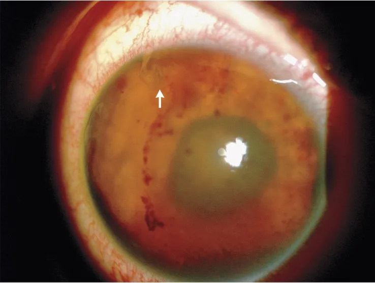
A 61-year-old woman experienced a strange side effect of her rheumatoid arthritis: a condition called "corneal melt," according to a 2014 report of the woman's case published in The New England Journal of Medicine.
The condition occurs when a person's immune system attacks the area of the eye next to the cornea, tearing the ocular tissue and allowing the iris, which sits behind the cornea, to "slip" out. (The cornea is the transparent layer of the eye that sits on top of the iris and the pupil.)
In the woman's case, both of her eyes were affected — a very rare occurrence, experts say.
Doctors can attempt to repair the eyes surgically, but that won't prevent the condition from happening again, according to the American Academy of Ophthalmology.
"Protruding" feature
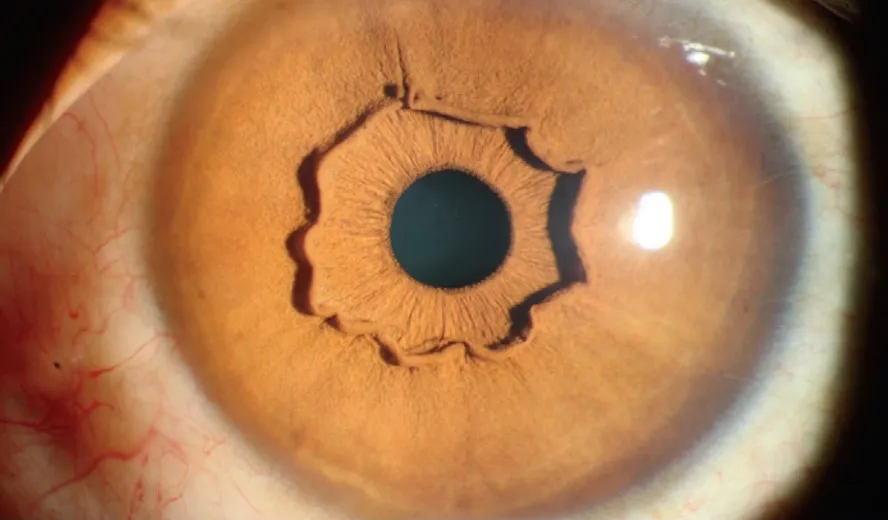
Doctors in China spotted an odd feature in a woman's eyes: a raised, rippled ring of tissue encircling the irises of both eyes.
The ring, called a "protruding iris collarette," isn't actually linked to any vision problems. Instead, it's a variation of a normally flat part of the eye called the iris collarette. In the woman's case, the doctors found that her eyes were healthy and her vision was normal, according to the report of the woman's case, published in March 2017 in The New England Journal of Medicine.
Although the protruding features in this woman's case were particularly pronounced, the condition isn't as rare as it may seem, Dr. Andrea Thau, the former president of the American Optometric Association, who was not involved in the case report, told Live Science in March 2017. Generally, the raised ring of tissue isn't as prominent as the one seen in this case, Thau said.
Limited diet leads to boy's vision loss

A highly restrictive diet that was limited to potatoes, pork, lamb, apples, cucumbers and Cheerios led to an 11-year-old boy's severe vision loss, according to a report of the boy's case.
None of those foods is a good source of vitamin A, and indeed, when doctors tested the boy's blood to measure levels of the vitamin, they found that he was severely deficient in vitamin A, according to the report, published in the journal JAMA Pediatrics in October 2017.
Vitamin A is essential for vision because it helps certain cells in the eyes function properly. Without enough of the vitamin, a person can develop severely dry eyes and a buildup of material on the outer covering of the eyes. Vitamin A deficiency can also lead to problems in the retina, which is home to light-sensing cells that make vision possible.
To treat the vitamin deficiency, doctors gave the boy "megadoses" of vitamin A intravenously. The appearance of his eyes improved significantly, but his vision may be permanently damaged, according to the report.
A hairy situation

It's not uncommon to get an eyelash caught in your eye, but for your eyeball to grow a hair of its own — well, that's a lot rarer.
A case report published in The New England Journal of Medicine in 2013 describes a 19-year-old Iranian man with a benign tumor on his right eye that sprouted several black hairs.
The man had the tumor, called a limbal dermoid, since birth, and it had gradually grown throughout his life. These rare tumors can contain tissue normally found in other parts of the body, such as hair follicles or sweat glands.
Though the tumors can lead to blurred vision, they usually don't cause dramatic vision problems because they don't cover the center of the eye. In the man's case, the tumor was surgically removed.
Flatworm wriggles through eye
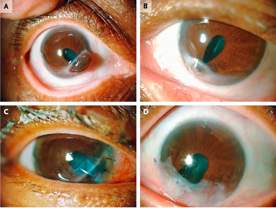
Doctors in Mexico spotted a flatworm wriggling in and out of a teen's right eyeball, according to a report of the teen's case.
By the time the teen sought medical attention, the worm, which the doctors were able to identify as a type of trematode or "fluke," had caused serious damage: The teen's right cornea was swollen and speckled with blood, and there were multiple holes in his iris due to the worm's movements.
The doctors were able to surgically remove the worm — though not all in one piece, according to the report, which was published in The New England Journal of Medicine in September 2017. During the surgery, the doctors needed to remove the lens of the teen's eye, along with the vitreous humor, which is the fluid that fills the eyeball.
Six months after the surgery, the doctors noted that the teen's vision in his right eye had not improved.
Gone fishin'

A tourist taking a dip in the Red Sea caught more than a few waves when he went swimming; he also ended up with a fish jaw stuck in his eyelid.
During the man's swim, he collided with a school of fish, according to a report of the man's case, which was published in The New England Journal of Medicine in 2015. However, at the time, the man didn't realize that a fish jaw was left behind in his eyelid.
The man developed a swollen and droopy eyelid, which didn't go away, according to the report. When he went to the doctor, an imaging test revealed that he had an area of inflammation called a granuloma in his eyelid. But when the doctors operated to remove this inflammation, they were surprised to find "two transparent tubular structures."
Later, a biologist identified the structures as the jawbones of a halfbeak fish, a species that is commonly found in shallow and coastal waters.
Under pressure
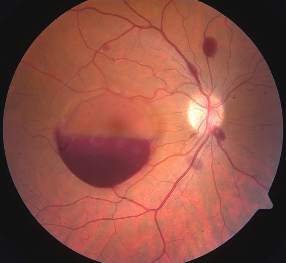
A 32-year-old man temporarily lost vision in one of his eyes after an intense session of handstand push-ups, according to a report of the man's case, published in the journal BMJ Case Reports in 2014.
The man told doctors that he lost vision in his right eye about 6 hours after his workout, which included doing push-ups in a handstand position.
When the doctors examined the man's eye, they found a large hemorrhage, or heavy bleeding, in front of his retina, also with several other smaller sites of bleeding throughout the eye. These spots of blood obscured the man's vision.
The man was diagnosed with a condition called valsalva retinopathy, which refers to internal bleeding that occurs after people attempt to exhale while holding their mouths closed and pinching their noses shut. This leads to a sudden increase in chest pressure, which causes the tiny capillaries in the eye to rupture.
Crash course
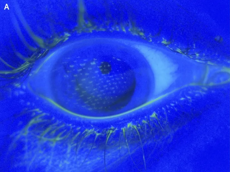
Airbags can save lives, but they can also leave behind some damage.
After a car accident, a 17-year-old in Michigan was left with an imprint of an airbag's canvas on her eyeballs; the airbag deployed before the teen could even blink, according to a report of the teen's case, published in The New England Journal of Medicine in 2014.
The teen went to the emergency room because she had pain and burning in her eyes. There, doctors used a special fluorescent dye that highlights scratches or tears on the cornea. The dye revealed an imprint of the nylon mesh pattern of the airbag cover in both eyes, according to the report. In addition to the imprints, the teen had a small tear in one of her eyes and some bleeding. However, all of the injuries healed in two weeks, the report said.
