Egyptian Mummy's Brain Imprint Preserved in 'Peculiar' Case

Get the world’s most fascinating discoveries delivered straight to your inbox.
You are now subscribed
Your newsletter sign-up was successful
Want to add more newsletters?

Delivered Daily
Daily Newsletter
Sign up for the latest discoveries, groundbreaking research and fascinating breakthroughs that impact you and the wider world direct to your inbox.

Once a week
Life's Little Mysteries
Feed your curiosity with an exclusive mystery every week, solved with science and delivered direct to your inbox before it's seen anywhere else.

Once a week
How It Works
Sign up to our free science & technology newsletter for your weekly fix of fascinating articles, quick quizzes, amazing images, and more

Delivered daily
Space.com Newsletter
Breaking space news, the latest updates on rocket launches, skywatching events and more!

Once a month
Watch This Space
Sign up to our monthly entertainment newsletter to keep up with all our coverage of the latest sci-fi and space movies, tv shows, games and books.

Once a week
Night Sky This Week
Discover this week's must-see night sky events, moon phases, and stunning astrophotos. Sign up for our skywatching newsletter and explore the universe with us!
Join the club
Get full access to premium articles, exclusive features and a growing list of member rewards.
An ancient Egyptian mummy is sparking new questions among archaeologists, because it has one very rare feature: The blood vessels surrounding the mummy's brain left imprints on the inside of the skull.
The researchers are trying to find what process could have led to the preservation of these extremely fragile structures.
The mummified body is that of a man who probably lived more than 2,000 years ago, sometime between the Late Period and the Ptolemaic Period (550 – 150 B.C.) of Egyptian history, the researchers said.
"This is the oldest case of mummified vascular prints" that has been found, study co-author Dr. Albert Isidro told Live Science in an email.
The mummy was recovered in 2010, along with more than 50 others in the Kom al-Ahmar/Sharuna necropolis in Egypt. [8 Grisly Archaeological Discoveries]
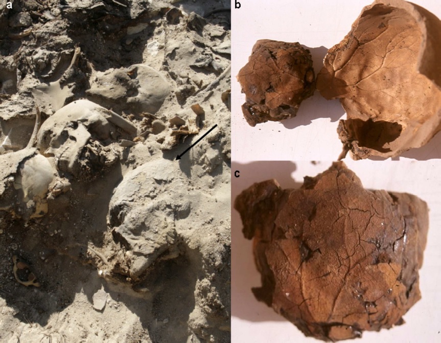
But unlike his neighbors in the field, the inside of this man's skull bore the imprints of his brain vessels, with "exquisite anatomical details," for centuries. The prints were cast into the layer of the preservative substances used during the mummification process to coat the inside of the skull.
The imprints appear to have been made by the blood vessels within the meninges, which is the membrane that covers the brain, the researchers said.
Get the world’s most fascinating discoveries delivered straight to your inbox.
"It is a truly remarkable finding and an interesting case," the researchers wrote in their report on the mummy, published Sept. 19 in the journal Cortex. To date, there have been only a few anecdotal reports of similar cases, they said.
The mummy, dubbed W19, was preserved using substances such as bitumen (a viscous oil) mixed with linen, the researchers found. The imprints of the vessels on the skull bone mirrored the prints on the mass of preservatives found within the skull, the researchers said. It was most likely a brain vessel called the middle meningeal artery that created the imprint, they said.
It is even possible that part of the man's actual meninges still remain there, in the outermost layer of the preservative mass, Isidro said. But the only way to know for sure would be to rehydrate the tissue and look for microscopic signs of the cells, he said.
During the mummification process that the Egyptians followed, the brain was removed, usually through the nose using wirelike instruments, and then the inside of the skull was cleaned and filled with preservative substances. It's unexpected for any brain tissue to remain intact after these procedures, Isidro said.
In this man, something peculiar must have happened when his body was being mummified, the researchers said.
"The conditions in this case must have been quite extraordinary," the researchers said. "We can speculate that something special happened in individual W19 just at the moment of bitumen insertion" into the skull.
But the researchers said they don't know what exactly happened. One possibility is that the general conditions, such as the temperature or the acidity, of the preservative, were different for W19 than for the other people whose mummies were found in the same necropolis, Isidro said.
Although brain tissue is rarely found in artificial mummies who undergo brain extraction, it has been discovered frequently in natural mummies who were preserved in just the right environment. For example, Europe's oldest mummy, Ötzi the Iceman, had some brain tissue preserved, which revealed information about the circumstances of his death.
Editor's note: This article was updated on Sept. 30, 2014 to include a new comment from the researchers about the possibility that the actual meninges remain in the preservative.
Email Bahar Gholipour. Follow Live Science @livescience, Facebook & Google+. Originally published on Live Science.

 Live Science Plus


