 Live Science Plus


You are now subscribed
Your newsletter sign-up was successful
Want to add more newsletters?

Delivered Daily
Daily Newsletter
Sign up for the latest discoveries, groundbreaking research and fascinating breakthroughs that impact you and the wider world direct to your inbox.

Once a week
Life's Little Mysteries
Feed your curiosity with an exclusive mystery every week, solved with science and delivered direct to your inbox before it's seen anywhere else.

Once a week
How It Works
Sign up to our free science & technology newsletter for your weekly fix of fascinating articles, quick quizzes, amazing images, and more

Delivered daily
Space.com Newsletter
Breaking space news, the latest updates on rocket launches, skywatching events and more!

Once a month
Watch This Space
Sign up to our monthly entertainment newsletter to keep up with all our coverage of the latest sci-fi and space movies, tv shows, games and books.

Once a week
Night Sky This Week
Discover this week's must-see night sky events, moon phases, and stunning astrophotos. Sign up for our skywatching newsletter and explore the universe with us!
Join the club
Get full access to premium articles, exclusive features and a growing list of member rewards.
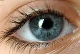
The first images ever made of retinas in living people reveal surprising variation from one person to the next. Yet somehow our perceptions don't vary as might be expected.
Imaging thousands of cells responsible for detecting color in the deepest layer of the eye, scientists found that our eyes are wired differently. Yet we all -- with the exception of the color blind -- identify colors similarly.
Inside the eye
The eye, responsible for receiving visual images, is wrapped in three layers of tissue [graphic]. The innermost layer, the retina, is responsible for sensing color and sending information to the brain.
Inside the Eye
The retina contains light receptors known as cones and rods. These receptors receive light, convert it to chemical energy, and activate the nerves that send messages to the brain. The rods are in charge of perceiving size, brightness and shape of images, whereas color vision and fine details are the responsibility of the cones.
On average, there are 7 million cones in the human retina, 64 percent of which are red, 32 percent green, and 2 percent blue, with each being sensitive to a slightly different region of the color spectrum. At least that's what scientists have been saying for years.
But the first complete imaging of the human retina, mapping the arrangement of the three types of cone photoreceptors, revealed something surprising about these numbers.
Big variation
The study found that people recognized colors in the same way. Yet the pictures of their retinas showed there is enormous variability, sometimes up to 40 times, in the relative number of green and red cones in the retina.
"[This] suggests that there is a compensatory mechanism in our brain that negates individual differences in the relative numbers of red and green cones that we observed," Joseph Carroll, a researcher at Center for Visual Science at University of Rochester and a collaborator of the study, told LiveScience.
The researchers used adaptive optics imaging, which uses a camera containing a corrective device that cancels the effects of the eye's imperfect optics on image quality, producing a high-resolution retinal picture.
Borrowing from astronomy
"Adaptive optics is a technique borrowed from astronomy where it is used to obtain sharp images of stars from telescopes on the ground," said David Williams, Director of Center for Visual Science at the University of Rochester. "All such telescopes suffer from blur due to the effects of turbulence in the Earth's atmosphere. In our case, optical defects in the cornea and lens of the eye blur images of the retina."
The measured defects were corrected using deformable mirrors, which bend and morph according each person's eye, before taking high magnification pictures of the eye. This allowed Williams and colleagues to see and map single cells such as the cones.
The researchers hope to use the same techniques to better understand various forms of color blindness and different kinds of retinal disease.
The findings were detailed in a recent issue of the Journal of Neuroscience.

