


Scientists Watch a Fish Think
For the first time, scientists have imaged the brain activity of a fish watching its prey.
Observing neural signals in real time offers an important glimpse into how brains perceive the outside world. In the new study, researchers developed a way to follow these signals in the brain of a zebrafish larva, using a sensitive fluorescent marker.
"It's a breakthrough," molecular and cell biologist Florian Engert of Harvard University, who was not involved in the study, told LiveScience. "No one else can look at neuronal activity with fluorescence microscopy in a freely swimming zebrafish larva" with such good resolution.
See-through heads
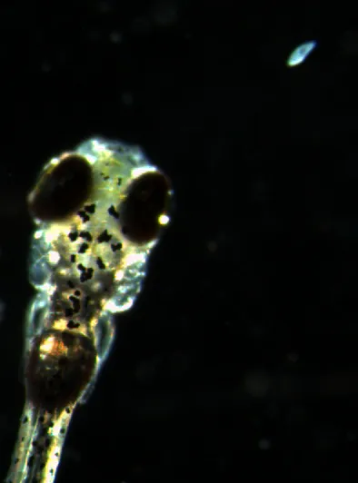
Zebrafish are widely used to study genetics and development in vertebrates. Their larvae are ideal for neuroimaging because they have translucent heads, and scientists can literally peer into their brains.
To see what was actually going on in those fish noggins, researchers developed a genetically engineered protein, called GCaMP7a, that lights up under a fluorescent microscope when neurons, or brain cells, fire. Transgenic zebrafish were bred to express this protein in a brain region called the optic tectum, which controls the movement of the eye when the animal sees something move in its environment.
In one experiment, the scientists imaged the brain of a transgenic fish larva as it watched a dot on a screen blinking on and off or moving back and forth. Under the microscope, signals flashed through the fish's brain, mirroring the movement of the dot. [See video of the fish's brain.]
Get the world’s most fascinating discoveries delivered straight to your inbox.
Next, a live paramecium — zebrafish prey — was placed in sight of an immobilized fish. Again, neural signals could be seen zipping around the fish's brain, tracking the paramecium's movement. No signals were detected when the paramecium was motionless, however.
Lastly, a paramecium was placed in a dish with a zebrafish larva that was allowed to swim freely, hunting its prey. The researchers mapped the fish's brain activity as it zeroed in on the paramecium and swam toward it.
Understanding brain behavior
The new approach will improve scientists' understanding of brain circuits involved in predatory behavior, the researchers report online today (Jan. 31) in the journal Current Biology. The system could be used to image other brain areas, too, allowing scientists to observe neurons involved in behavior and locomotion.
Previously, scientists had been able to image single-cell brain activity in zebrafish, but this study was the first to do it in a freely swimming fish perceiving a natural object. "The technology for studying zebrafish is moving fast," said neuroscientist Joseph Fetcho in an email to LiveScience. Fetcho did some of the earlier imaging work but was not involved in the new study.
The closer one can get to revealing the patterns of neuronal activity in a freely behaving animal, the more likely the patterns will represent those that drive natural behavior, Fetcho said.
Follow LiveScience on Twitter @livescience. We're also on Facebook & Google+.
