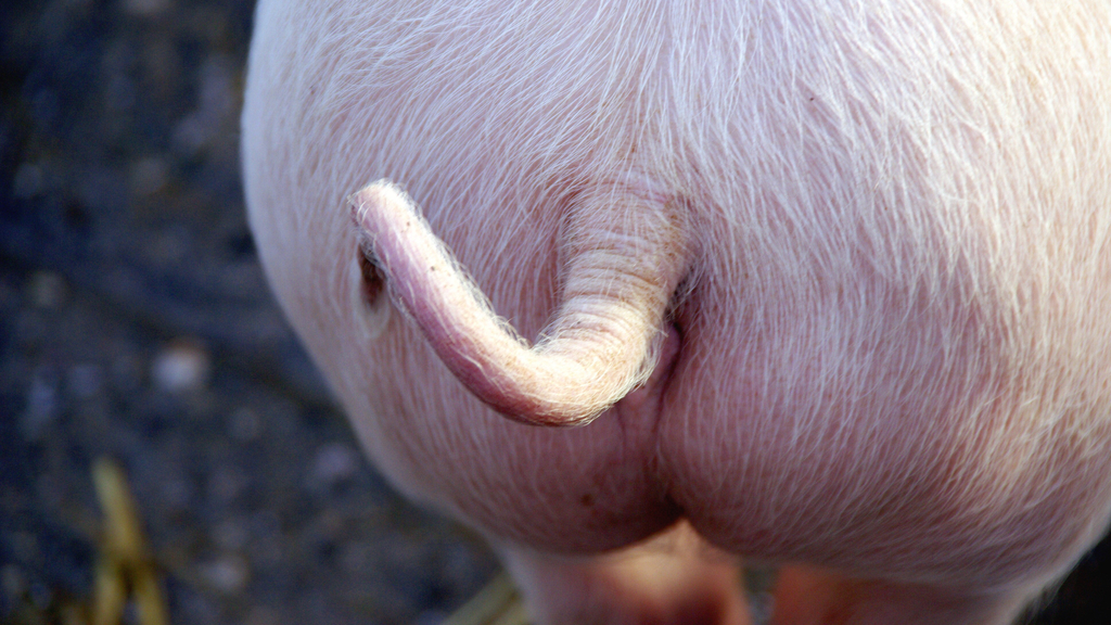
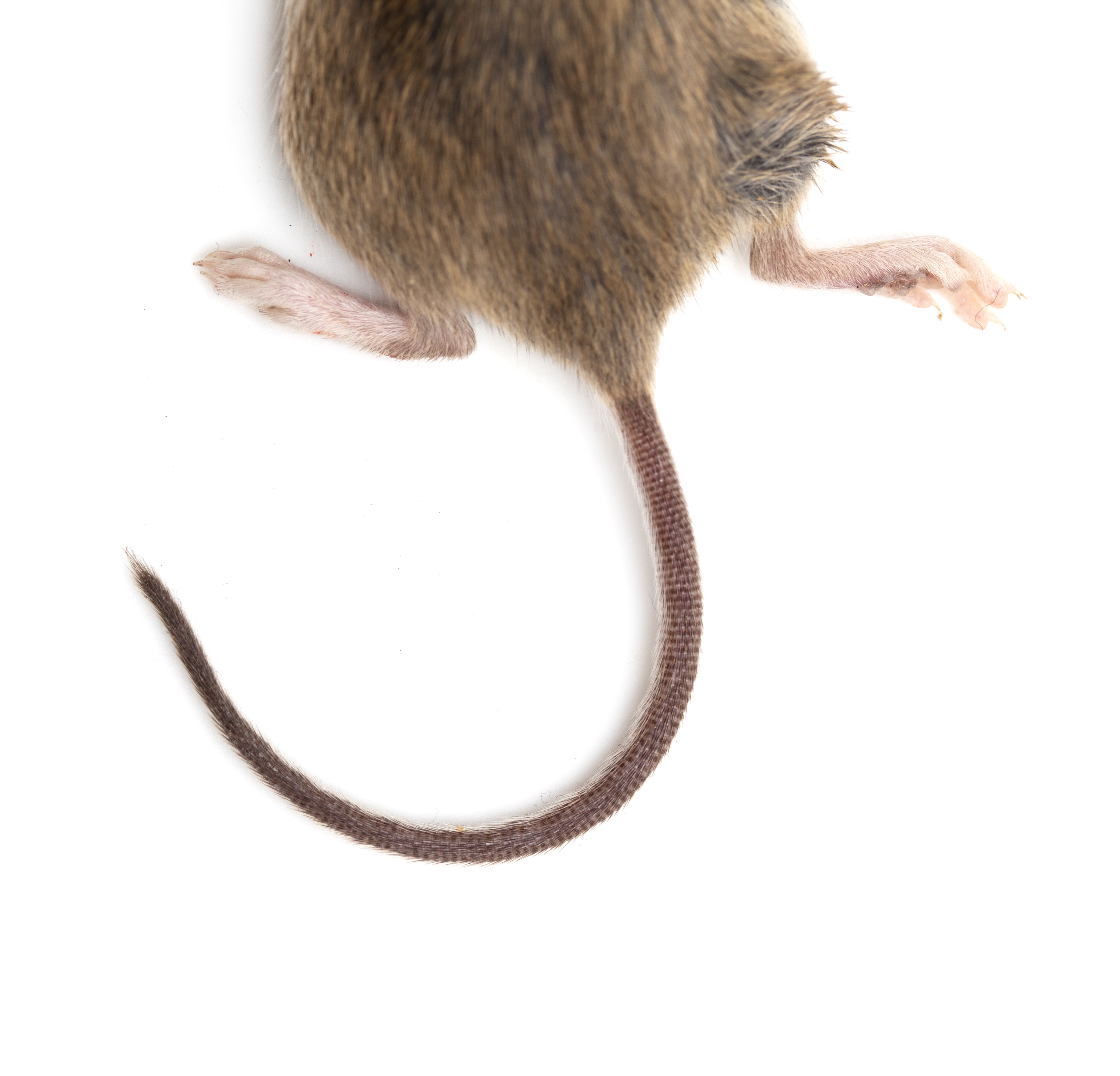
Pigs can breathe through their butts. Can humans?

Mice, rats and pigs all share a secret superpower: They can all use their intestines to breathe, and scientists discovered this by pumping oxygen up the animals' butts.
Why run such experiments, you ask? The research team wanted to find a potential alternative to mechanical ventilation, a medical treatment where a machine pushes air into a patient's lungs through the windpipe. Ventilators deliver oxygen to the lungs and help remove carbon dioxide from the blood, but the machines aren't always available.
Early in the COVID-19 pandemic, for example, hospitals faced a severe shortage of ventilators, The New York Times reported. Although doctors can also use a technique called extracorporeal membrane oxygenation (ECMO), where blood is pumped out of the body and reoxygenated with a machine, the procedure carries inherent risks, such as bleeding and blood clots; and it's often less readily available than ventilators, according to Mayo Clinic.
Related: The 10 weirdest medical cases in the animal kingdom
In search of another solution, the study authors drew inspiration from aquatic animals like sea cucumbers and freshwater fish called loaches (Misgumus anguillicandatus), which use their intestines for respiration. It was unclear whether mammals have similar capabilities, although some scientists attempted to answer that question in the 1950s and 1960s.
"We initially looked at a mouse model system to see if we could deliver oxygen gas intra-anusly," said senior author Dr. Takanori Takebe, a professor at the Tokyo Medical and Dental University and a director at the Center for Stem Cell and Organoid Research and Medicine at Cincinnati Children's Hospital Medical Center.
"Every time we performed experiments, we were quite surprised," Takebe told Live Science.
Get the world’s most fascinating discoveries delivered straight to your inbox.
Without intestinal ventilation, mice placed in a low-oxygen environment survived for only about 11 minutes; with ventilation into their anuses, 75% survived for 50 minutes, thanks to an infusion of oxygen that reached their hearts. The team then tried using oxygenated liquid, rather than gas, in mice, rats and pigs, and they found similarly promising results. The team noted that more work still needs to be done to see if the approach is safe and effective in humans, according to a paper on their findings published May 14 in the journal Med.
"The pandemic has highlighted the need to expand options for ventilation and oxygenation in critical illness, and this niche will persist even as the pandemic subsides," as there will be times when mechanical ventilation is unavailable or inadequate on its own, Dr. Caleb Kelly, a clinical fellow and physician-scientist at Yale School of Medicine, wrote in a commentary of the study. If, after further evaluation, intestinal ventilation eventually becomes common practice in intensive care units, this new study "will be marked by historians as a key scientific contribution," he wrote.
That said, a research group in Russia has already explored the idea of using intestinal ventilation in human patients and first conducted a clinical trial of the method in 2014, as described in the European Journal of Anaesthesiology. The same group, led by Dr. Vadim Mazurok, a professor and head of the anaesthesiology and intensive care department at the Almazov National Medical Research Centre, has also patented methods and equipment for delivering oxygen gas into the intestines. Takebe and his team will likely focus on using oxygenated liquid in human patients in their future clinical trials, but this previous work by Mazurok and his colleagues sets a precedent for the approach.
Getting familiar with loach, mouse and pig guts
Before starting their experiments in rodents, Takebe and his colleagues got very familiar with loach guts. The fish take in oxygen mostly through their gills, but occasionally, when exposed to low-oxygen conditions, loaches instead use a portion of their intestines for gas exchange, Takebe said. In fact, in response to the lack of oxygen, the structure of gut tissues near the anus changes such that the density of nearby blood vessels increases and secretion of fluids related to digestion decreases.
These subtle changes allow loaches to "suck up the oxygen more efficiently," Takebe said. In addition, the outermost lining of the loach gut — the epithelium — is very thin, meaning oxygen can easily permeate the tissue to reach the blood vessels beneath, he added. To simulate this structure in their mouse models, the team thinned out the gut epithelium of the rodents using chemicals and various mechanical procedures.
They then placed the mice under extremely low-oxygen conditions and used a tube to pump oxygen gas up the animals' bums and into their large intestines.
Related: 8 bizarre animal surprises from 'True or Poo' — Can you tell fact from myth?
Compared with mice whose gut epithelium had not been thinned, the mice with thin epitheliums survived significantly longer in the experiment — with most surviving 50 minutes as compared with about 18 minutes. Again, mice not given any oxygen only survived for about 11 minutes. In addition to surviving longer, the group with thinned-out gut linings showed signs that they were no longer starved for oxygen; they stopped gasping for air or showing signs of cardiac arrest, and the oxygen pressure in their major blood vessels improved.
Although this initial experiment suggested that oxygen could pass through the intestine and into circulation, thinning out the gut epithelium would likely not be feasible in human patients, Takebe said.
Particularly in critically ill patients, "I think additional damage to the gut would be really dangerous, for the treatment perspective," Takebe said. But "over the course of the experiments, we realized that even the intact gut has some, not really efficient, but some capacity to exchange the gas," he noted, meaning there may be a way to introduce oxygen through the gut without first thinning out the tissues.
So in another experiment, rather than using oxygen gas, the team tried perfluorodecalin (PFD), a liquid fluorocarbon that can be infused with a large amount of oxygen. The liquid is already used in people, such as for use in the lungs of infants with severe respiratory distress, the authors noted in their report.

The liquid also acts as a surfactant — a substance that reduces surface tension; since a surfactant lines the air sacs of the lungs and helps boost gas exchange in the organ, PFD may fulfill a similar purpose in the intestines, Takebe said.
Much like in the oxygen-gas experiments, the oxygenated PFD rescued mice from the effects of being placed in a low-oxygen chamber, enabling the rodents to meander about their cage more than mice not given the treatment. After just one injection of 0.03 ounces (1 milliliter) of the liquid, the rodents' improvements persisted for about 60 minutes.
"We are not quite sure why this improvement is persisting much longer than the original expectations," Takebe noted, as the authors expected the effects to wear off in just a couple minutes. "But the observation is really reproducible and very robust."
Related: Gasp! 11 surprising facts about the respiratory system
The team then moved on to a pig model of respiratory failure, where they placed pigs on ventilators and only provided a low level of oxygen and then injected PDF into the pigs' posteriors with a long tube. Compared with pigs not given the PFD treatment, pigs given PFD improved in terms of the oxygen saturation of their blood, and the color and warmth returned to their skin. A 13.5 oz (400 ml) infusion sustained these improvements for about 18 to 19 minutes, and the team found that they could give additional doses to the pigs without noticeable side effects.
The team also tested the safety of repeat dosing in rats and found that, while their oxygen levels rose, the animals showed no notable side effects, markers of organ damage or stray PFD lingering in their cells.
Following this success in animal models, Takebe said that his team hopes to start a clinical trial of the treatment in humans sometime next year. They would likely start by testing the safety of the approach in healthy volunteers and beginning to work out what dose levels would be reasonable, he said. However, to make the jump from animals to human patients, the team will need to address a number of critical questions.
For instance, the treatment could potentially stimulate the vagus nerve — a long nerve that connects the gut and brain — so trial organizers should likely be on the lookout for side effects like falling blood pressure or fainting, Takebe noted. Also, the lower gut contains relatively little oxygen compared with other organs in the body, he added. The community of bacteria and viruses that live in the gut are adapted to these low-oxygen conditions, and a sudden infusion of oxygen might disrupt those microbes, he said.
"The consequence of reversing this so-called 'physiologic hypoxia' is unknown," Kelly noted in his commentary, echoing Takebe's sentiments. In humans, it will be important to determine how many doses of oxygenated liquid could be safely administered into the gut without causing unintended changes to the intestinal environment, he wrote.
In addition, the animal models in the study don't fully reflect what critically ill patients experience during respiratory failure, a condition that often coincides with infection, inflammation and low blood flow, Kelly noted. So there may be additional factors to consider in critically ill patients that weren't relevant in rodents and pigs. And depending on a given patient's condition, they may need a higher or lower dose of PFD — all of these fine details will need to be carefully assessed in future trials, Takebe said.
Editor's note: This story was updated on May 19 to note the previous work of Dr. Vadim Mazurok and his colleagues, who have patented methods of intestinal ventilation in human patients. The original story was published on May 14.
Originally published on Live Science.

Nicoletta Lanese is the health channel editor at Live Science and was previously a news editor and staff writer at the site. She holds a graduate certificate in science communication from UC Santa Cruz and degrees in neuroscience and dance from the University of Florida. Her work has appeared in The Scientist, Science News, the Mercury News, Mongabay and Stanford Medicine Magazine, among other outlets. Based in NYC, she also remains heavily involved in dance and performs in local choreographers' work.
