
 Live Science Plus


Hind Sight: Blind Tadpoles See Via Eyes in Tails

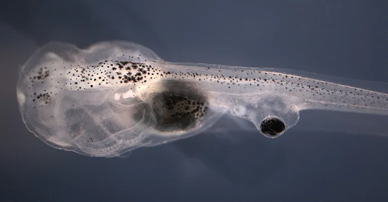
If you've ever wished you had eyes on the back of your head, meet the amphibian with eyes on its butt: Researchers have enabled tadpoles to see through eyes grafted onto their tails.
The project represents a promising step forward in the world of organ transplants and regenerative medicine, the researchers said.
The scientists grafted eye tissue onto blind tadpoles' tails, then dosed the animals with a drug that promotes neural development, building new connections between the implanted eyes and the tadpoles' nervous systems. In experiments, the tadpoles could distinguish between colors and could follow rotating patterns, even though the eyes were not connected to the animals' brains the same way that natural eyes would be, the study said.

"The fact that the grafted eyes in our model system could transmit visual information, even when direct connections to the brain were absent, suggests the central nervous system contains a remarkable ability to adapt to changes both in function and connectivity," study lead author Douglas Blackiston, a postdoctoral researcher at Tufts University in Massachusetts, said in a statement. [Through Their Eyes: Awesome Photos Show How Animals See the World]
Tadpoles are commonly used for studies of tissue grafts, because from the earliest stages of the animals' development, they can be manipulated genetically, surgically and with drugs, study co-author Michael Levin, a professor of biology at Tufts and the director of the university's Center for Regenerative and Developmental Biology, told Live Science in an email.
This makes tadpoles suitable for many lines of research — "everything from birth defects to immunology to neurological disease," Levin explained.
In an earlier study, Levin and Blackiston surgically removed tadpoles' developing eyes and grew new eyes on the animals' tails and torsos. These grafted eyes were identical in size and shape to eyes that the animals would have developed normally, and could be grown at just about any place on the tadpoles' lower bodies except at the very end of their tails, the scientists reported.
Get the world’s most fascinating discoveries delivered straight to your inbox.
However, those new eyes didn't work so well, the scientists said. The eyes showed a only limited sensitivity to light, and the researchers questioned how performance might be improved, Levin said.
Rear view
In the new study, the researchers added a drug that stimulates neural growth to their technique. The drug, called Zolmitriptan, affects serotonin levels and is approved for treating people who have migraines.
The researchers found that the drug enhanced the development of neural connections in tadpoles. Although the grafted optic tissue did not connect directly to the brain, as normal eyes would do, new connections were formed that provided the brain with sensory input, even when the optical signals originated from an unexpected place — the tadpoles' rear ends, according to the study.
The scientists then tested the tadpoles' ability to use their new eyes to detect the difference between red and blue. Tadpoles were trained in chambers illuminated by red and blue lights, learning to associate the color red with receiving a mild electric shock. They were then presented with the opportunity to enter a blue space or a red one. Of the tadpoles that had received the drug treatment, 29 percent showed preference for blue, compared to 11 percent of tadpoles that had new eyes, but had received no neurotransmitter enhancer.
Another test tasked tadpole in a petri dish with following the direction of rotating optical patterns that were visible on the screen of a monitor placed underneath their dish. Again, tadpoles that received the drug performed better, with 57 percent swimming in the same direction as the moving pattern, compared to 32 percent of the tadpoles that did not get the drug.
Growth opportunity
Restoring function in grafted tissues or organs depends on those tissues making connections to the neural networks that control them. The new results hint that the brain is adaptable enough to process input from unexpected locations and along unfamiliar pathways, Levin explained.
"The brain is remarkably plastic," Levin told Live Science in an email. "A tadpole brain, evolving for eons to expect visual input from the standard eye locations, has absolutely no problem picking up visual data from a weird new location on its back. This means that the brain can map its behavioral programs onto new body architectures, and this will be useful in the future, to improve not only transplants addressing regenerative medicine needs, but also perhaps in sensory/motor augmentation."
Or, to put it another way, "The body can change drastically, and the brain will follow," Levin said.
The findings were published online today (March 30) in the journal npj Regenerative Medicine.
Original article on Live Science.

Mindy Weisberger is a science journalist and author of "Rise of the Zombie Bugs: The Surprising Science of Parasitic Mind-Control" (Hopkins Press). She formerly edited for Scholastic and was a channel editor and senior writer for Live Science. She has reported on general science, covering climate change, paleontology, biology and space. Mindy studied film at Columbia University; prior to LS, she produced, wrote and directed media for the American Museum of Natural History in NYC. Her videos about dinosaurs, astrophysics, biodiversity and evolution appear in museums and science centers worldwide, earning awards such as the CINE Golden Eagle and the Communicator Award of Excellence. Her writing has also appeared in Scientific American, The Washington Post, How It Works Magazine and CNN.