It's quick and easy to access Live Science Plus, simply enter your email below. We'll send you a confirmation and sign you up for our daily newsletter, keeping you up to date with the latest science news.
Someday, a mother-to-be may be able to put on a virtual reality headset and get a clear, 360-degree look at her own fetus in the womb. This scenario may sound like science fiction, but, according to a team of researchers from Brazil, it may be widely available in only a year.
Although 3D imaging of fetuses is already available to a great extent — and in fact there are portrait studios that offer it — the images that are obtained from current techniques are static, and still rather unclear.
With the new virtual reality technique, the images of a fetus are clearer and can be rotated 360 degrees. The researchers said that they hope that these enhanced fetal models are the next step in not only allowing parents to visualize their future children, but also in helping researchers to better understand fetal anatomy. [7 Baby Myths Debunked]
In their research, the researchers were able to use the technique to visualize and make 3D models of 25 fetuses. There were two cases in which the technique didn't work. In those, the levels of amniotic fluid were too low for the researchers to get images of the fetus that were high enough in their resolution to make the 3D model, Werner told Live Science.
But in the cases where the technique worked, "we found these images more real, and the possibility that we can see in 360 degrees presents us with a greater interaction with the exam," said study co-author Dr. Heron Werner Jr., who is from a company called Clinical Diagnostic Imaging that is based in Rio de Janeiro. Heron and his colleagues recently presented the technique at the annual meeting of the Radiological Society of North America.
The new technique involves first producing 3D models of a fetus using MRI and ultrasound techniques, or a combination of the two. For a pregnant woman, this would mean undergoing an imaging exam that is similar to a regular obstetric ultrasound, or MRI. But afterward, the researchers would use frames of these images, in sequence, to begin to make a 3D model of the fetus, the researchers said. [5 Fascinating Facts About Fetal Ultrasounds]
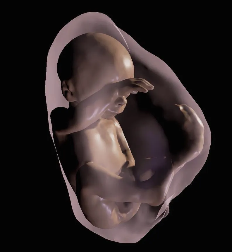
This is a close-up of fetus at 26 weeks.
(Image credit: Radiological Society of North America)
From there, a physician could choose to focus only on specific body parts of the fetus for the reconstruction. Finally, the parents-to-be could view the final image — which can include the inside of the womb, the umbilical cord and the placenta along with the fetus — through a virtual reality device.
Sign up for the Live Science daily newsletter now
Get the world’s most fascinating discoveries delivered straight to your inbox.
Werner and his colleagues used Oculus Rift 2, a virtual reality headset, in their research. They found that women could not only experience what it would look like if they were flying through and around their fetus by merely looking around, but also they could hear the fetal heartbeat, by way of the ultrasound.
Aside from its potential to give parents an extra-special sneak peek at their bundle of joy, this imaging technology could provide new options for evaluating the health and development of a fetus, the researchers said. The scan allows users to see all of the internal structures as well as external features of the fetus. For example, the researchers said, a doctor could zoom through the entire length of a fetus’s airway to look for masses that could block it and to better determine delivery options.
In this research, the scans did not reveal any health conditions in the fetuses that were not already known. But Werner said that the technique did improve the ability of multidisciplinary health care teams to work together and talk with family members about potential health problems.
The new technology could also help with planning pre- and postnatal surgeries, he said.