At just two-and-a-half hours old, a teensy fruit-fly embryo is bustling with activity, and now researchers have captured this development in a 3D video showing a young embryo grow into a 20-hour-old larva.
"This video shows a fruit fly embryo from when it was about two-and-a-half hours old until it walked away from the microscope as a larva, 20 hours later," Lars Hufnagel, from the European Molecular Biology Laboratory (EMBL) in Heidelberg, Germany, said in a statement. "It shows all the hallmarks of fruit-fly embryonic development in three dimensions."
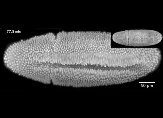
The video reveals what looks like a bloblike organism covered with tiny projections, which are actually various cells and cell parts, moving across its body. For instance, the cells on the embryo's belly dive inward to form the so-called ventral furrow, or an indentation that results in the formation of a tube inside the embryo marking the start of the gastrulation stage of development; other cells move around the embryo's rear end to its back in a process called convergent extension. Later in the video, an opening appears on the embryo's back before surrounding cells close this gap (a process called dorsal closure). [Science Meets Art in Stunning Images]
Latest Videos From
The new microscope, called Multi-View SPIM (MuVi-SPIM) shines a thin sheet of light on the embryo, illuminating one layer of the embryo at a time to prevent light damage. It takes four full images from different angles, which means scientists don't have to rotate the sample. The images are then merged to create a three-dimensional look at the sample, in this case a fruit-fly embryo. This whole process takes just seconds, so the microscope can repeat the process quickly.
As such, the different images that make up the video are taken in such rapid succession that very little has changed in the embryo from one frame to the next; in that way, scientists can be sure of the location of each cell, or even structures inside cells, and track it throughout the video.
In this new study, detailed this week in the journal Nature Methods, Hufnagel and colleagues recorded the movements of every cell nucleus in the embryo not only during the later developmental stages, but also throughout the first three hours of the embryo's life, when nuclei divide very rapidly.
In the future, the scientists hope to use their new microscope to investigate how organs and tissues form in the fruit fly and other organisms.
Follow LiveScience for the latest in science news and discoveries on Twitter @livescience and on Facebook.