Image Gallery: Science Meets Art
Get the world’s most fascinating discoveries delivered straight to your inbox.
You are now subscribed
Your newsletter sign-up was successful
Want to add more newsletters?

Delivered Daily
Daily Newsletter
Sign up for the latest discoveries, groundbreaking research and fascinating breakthroughs that impact you and the wider world direct to your inbox.

Once a week
Life's Little Mysteries
Feed your curiosity with an exclusive mystery every week, solved with science and delivered direct to your inbox before it's seen anywhere else.

Once a week
How It Works
Sign up to our free science & technology newsletter for your weekly fix of fascinating articles, quick quizzes, amazing images, and more

Delivered daily
Space.com Newsletter
Breaking space news, the latest updates on rocket launches, skywatching events and more!
Once a month
Watch This Space
Sign up to our monthly entertainment newsletter to keep up with all our coverage of the latest sci-fi and space movies, tv shows, games and books.

Once a week
Night Sky This Week
Discover this week's must-see night sky events, moon phases, and stunning astrophotos. Sign up for our skywatching newsletter and explore the universe with us!
Join the club
Get full access to premium articles, exclusive features and a growing list of member rewards.
Where Art & Science Intersect
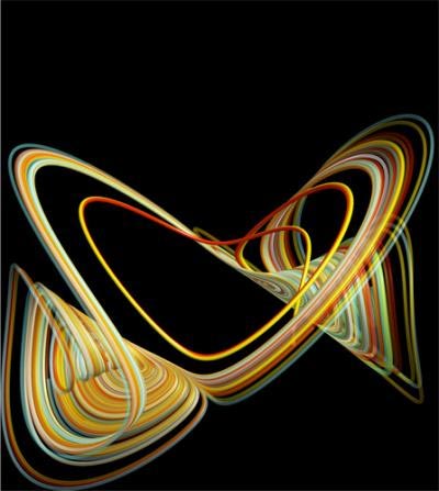
Princeton University's annual Art of Science exhibition explores the interplay between science and art, with each piece in the exhibit revealing those moments of discovery when what you perceive suddenly becomes more than the sum of its parts. In 2011, the fifth year of the competition, 168 pieces of art were submitted from 20 university departments, with 56 works chosen for the exhibition, each meant to fit with the year's theme of "intelligent design." (Shown above, an image created from a model illustrating the reversals of the Earth's magnetic field; these polarity reversals have occurred several times over the past 160 million years.)
Tree Art

Snagging second place, an image of a tree cut into smaller rectangular pieces. "As part of my research I am designing intelligent image decomposition algorithms that split an image into sub-images in a way that best captures important image structure," Zhen James Xiang said in a statement. "Natural images have structure. Understanding this structure and being able to decompose an image in a way that respects this structure is an important aspect of computational image processing."
To visualize how Xiang's decomposition algorithm works, he developed computer code that displays the resulting dyadic tree. The input image has been automatically cut into local rectangular pieces in a way carefully designed to achieve a useful global optimality.
For clarity, only a partial decomposition of the input image has been shown, reminding us of the inspirations we receive from nature: that harmony is required between division and unity, Xiang said.
Making Planets
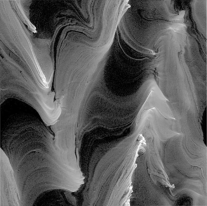
Planets form from the coagulation of tiny solid particles (dust) in a gaseous protoplanetary disk, requiring growth over 40 orders of magnitude in particle mass. A crucial stage in planet formation involves making kilometer-sized planetesimals from millimeter- to centimeter-sized pebbles. This image illustrates this process: aerodynamical interactions between the gas and the pebbles collect the latter into very dense clumps (bright regions), almost as if by design. In turn, these clumps become planetesimals, the building blocks of planets.
Artsy Arsenic
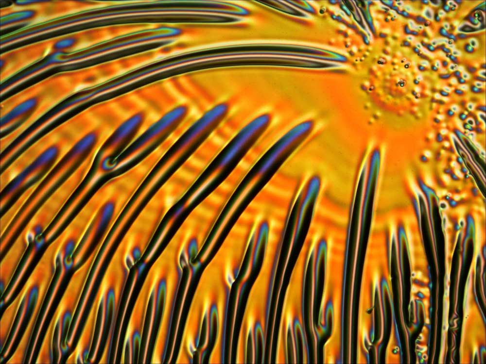
Arsenic sulphide dissolved in a solution displays colorful random patterns after being spin-coated and baked on a chrome-evaporated glass slide.
Baby Dragon
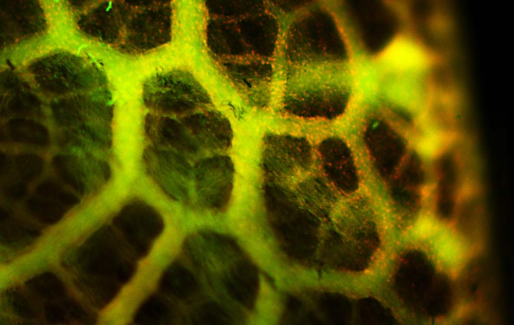
This is a detail of an immunofluorescence image of the surface of the lung of a bearded dragon embryo (Pogona vitticeps). Nuclei are stained red and the actin cytoskeleton, which helps cell movement, is stained green. The image reveals a nested hierarchy of tubes designed for effective gas exchange, which develops in the embryo even before the animal breathes air.
Electrified Crystals

Piezoelectric nanostructures, or those that produce an electric charge when a mechanical stress, such as squeezing or stretching, is applied could provide a clean alternative energy source. The crystal structures in this image were formed when the material was placed under high temperature and pressure.
Standing Embryos
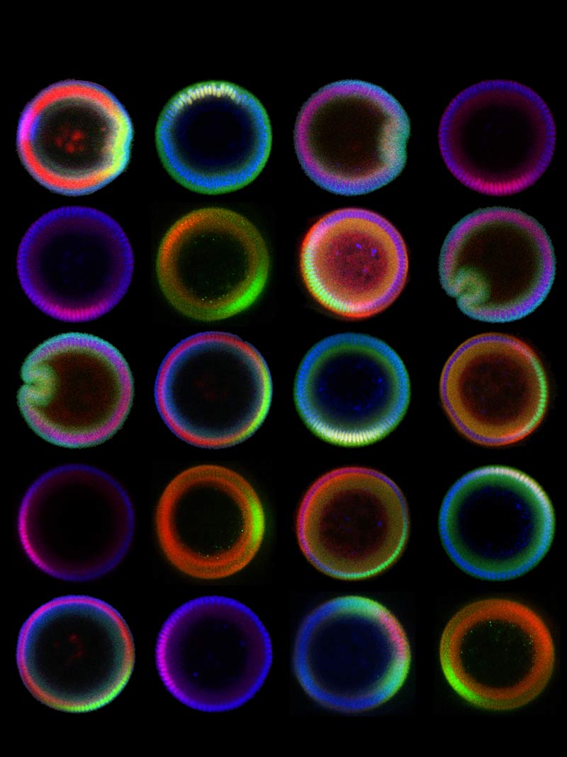
These vertical cross-sectional images of embryos of the common fruit fly (Drosophila melanogaster) are stained with antibodies in order to visualize molecules that subdivide the embryo into three tissue types: muscle, nervous system and skin.
Obtaining such images is an engineering challenge since it requires upright positioning of a tiny embryo, which is shaped like an ellipsis and just a half-millimeter long.
In collaboration with Lu lab at Georgia Tech, Princeton scientists have developed a device to trap and orient a large number of embryos vertically. The technique can be used to study embryos and, eventually, to understand the processes that drive the development of the embryo.
Get the world’s most fascinating discoveries delivered straight to your inbox.
Blurry Butterflies
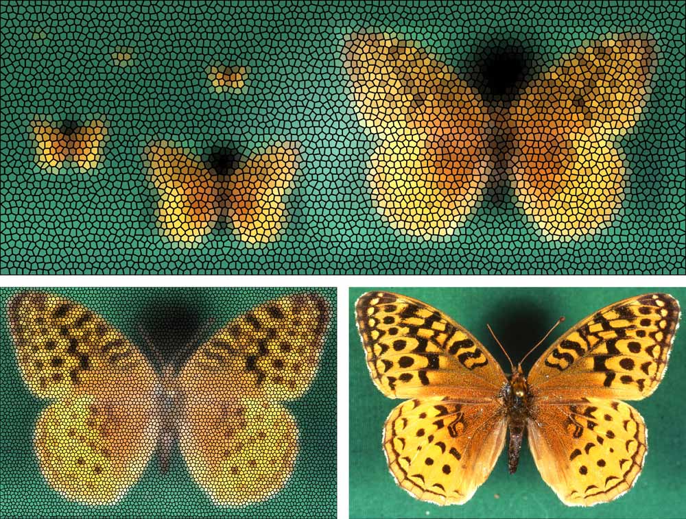
A simulated compound-eye view shows how a Great Spangled Fritillary Butterfly sees another Great Spangled Fritillary Butterfly from different distances (top) — (from upper left to right) 14.1 feet (4.3 meters), 6.9 ft. (2.1 m), 3.9 ft. (1.2 m), 2.3 ft. (0.71 m), 1.2 ft. (0.38 m), and finally the largest image you see on the top right, at a distance of only 0.59 ft. (0.18 m or 18 centimeters).
Below left is a simulated view at just (7 centimeters) compared with the original photograph (right). At 18 centimeters a striking phenomenon occurs: if the "eye" or the subject moves slightly, large portions of the field of view seem to flash between all orange and all black. It may be more than coincidence that 18 centimeters is about the typical courtship distance for this species.
Eye Tricks
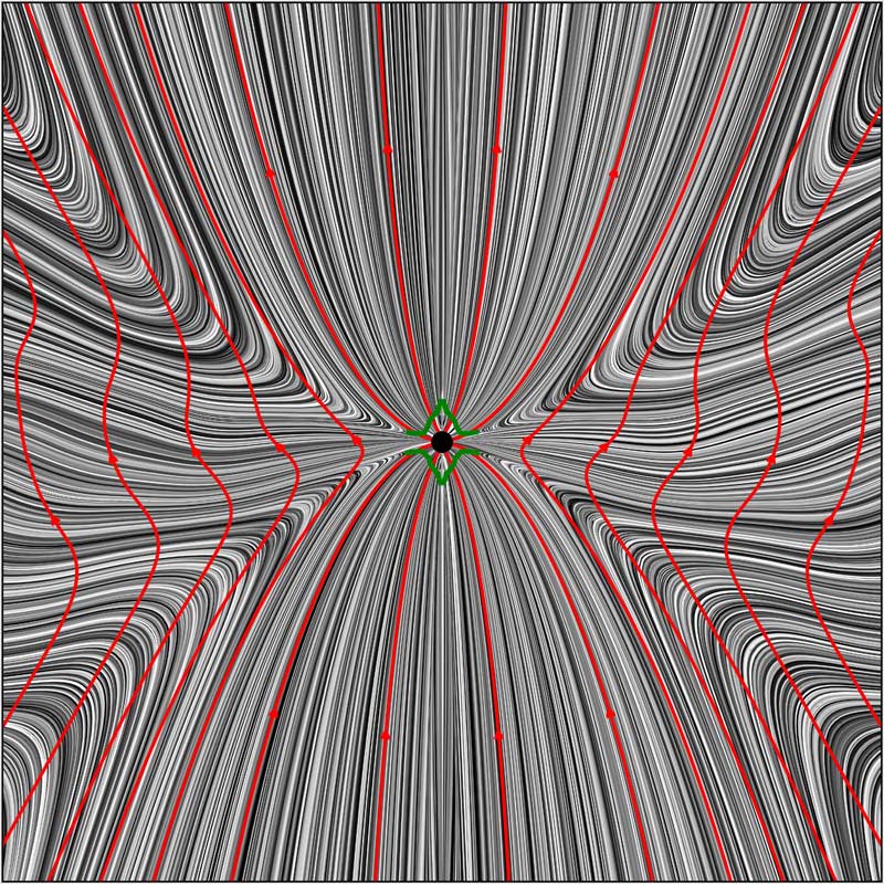
Simulated black-hole outflow powered by magnetic fields, which obstruct matter infall onto the hole. The black dot in the center shows the black hole horizon; gray lines show matter streamlines; red lines show magnetic field lines; and green lines show the boundary between the inflow and outflow.
Schooling Fish
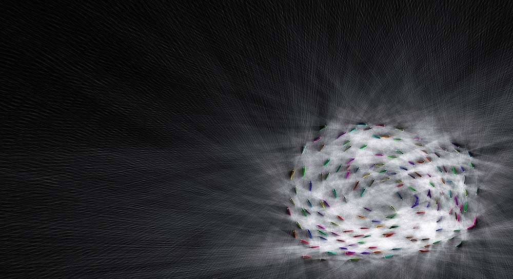
This image is a visualization of 150 fish (Notemigonus crysoleucas) free-swimming in a shallow 2.1 x 1.2 meter tank. It shows the recorded position of the body and eyes of each fish in the school for one frame of video.
Superimposed is a two-dimensional approximation of the field-of-view for each eye of each fish, shown as white rays cast outwards from the eye. Rays are terminated when they collide with another individual or the boundary of the arena.
This rough estimate of what each fish can see from its vantage point in the school is helpful for determining what information an individual has about its neighbors and environment at a given moment. This, in turn, allows scientists to study how information about a stimulus, such as a predator or food, may propagate through a group, changing the configuration of the group itself.
 Live Science Plus


