New Insight into How Eyes Form in Embryos
Get the world’s most fascinating discoveries delivered straight to your inbox.
You are now subscribed
Your newsletter sign-up was successful
Want to add more newsletters?
Delivered Daily
Daily Newsletter
Sign up for the latest discoveries, groundbreaking research and fascinating breakthroughs that impact you and the wider world direct to your inbox.
Once a week
Life's Little Mysteries
Feed your curiosity with an exclusive mystery every week, solved with science and delivered direct to your inbox before it's seen anywhere else.
Once a week
How It Works
Sign up to our free science & technology newsletter for your weekly fix of fascinating articles, quick quizzes, amazing images, and more
Delivered daily
Space.com Newsletter
Breaking space news, the latest updates on rocket launches, skywatching events and more!
Once a month
Watch This Space
Sign up to our monthly entertainment newsletter to keep up with all our coverage of the latest sci-fi and space movies, tv shows, games and books.
Once a week
Night Sky This Week
Discover this week's must-see night sky events, moon phases, and stunning astrophotos. Sign up for our skywatching newsletter and explore the universe with us!
Join the club
Get full access to premium articles, exclusive features and a growing list of member rewards.
Like tiny automatons, the cells that form a fish embryo's eyes are chemically programmed to individually amass at the site where the eyes will develop, according to a new study that contradicts traditional views of how organs develop before birth.
The study was done only on fish eyes and might or might not apply to humans.
"We think organs might be forming by the individual movement of cells," says Martina Rembold, of the European Molecular Biology Laboratory and the study's lead author.
Scientists previously thought that the eyes formed as cells at the sides of the tube-like structure that eventually forms the embryo's head and brain collectively bulged out—like blowing up the ears of a Mickey Mouse balloon, Rembold said. But she and her colleagues learned that the cells actually independently travel from the center of the tube out to the site of eye formation.
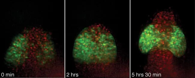
In 2001, Rembold discovered that only cells that manufacture a protein called Rx3 were involved in eye formation. For the new study, she followed the movements of these cells in a Medaka fish embryo by tagging them with a fluorescent marker. She used a microscope and camera to take pictures every 2 minutes for 10 hours. From that she created a stop animation movie [image] that allowed her "to follow all the individual cells throughout their development," she said.
From the starting point of the cells' journey, the protein attracts them to chemical signposts that guide them to the point where the eyes form. If the protein is not present in the cells, the cells won't migrate and the embryo's eyes won't develop.
Rembold says that other organs, like the heart, might form in a similar way, with cells individually traveling from other areas of the embryo.
Get the world’s most fascinating discoveries delivered straight to your inbox.
"It could be a universal mechanism," she said. But further research is needed to see whether the mechanism applies to other organs and other animals.

Andrea Thompson is an associate editor at Scientific American, where she covers sustainability, energy and the environment. Prior to that, she was a senior writer covering climate science at Climate Central and a reporter and editor at Live Science, where she primarily covered Earth science and the environment. She holds a graduate degree in science health and environmental reporting from New York University, as well as a bachelor of science and and masters of science in atmospheric chemistry from the Georgia Institute of Technology.
