Researchers have developed a three-dimensional model that catches a virus in the act of infecting a host cell.
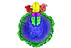
The model is one of the most detailed images of the tiny infectious agents ever produced. It shows a virus called Epsilon 15 poised to inject a long coil of DNA down a cylinder into a Salmonella bacterium.
Viruses infect cells by injecting their genetic material, either DNA or RNA, into host cells. Until now, however, scientists have never actually seen this process occur because viruses are so small.
Latest Videos From
The researchers photographed about 15,000 frozen viruses and fed the images into a computer. The images were then used to construct a 3D-model based on common features.
A team of researchers from Northwestern University also recently created a 3D virus model using another technique, called X-ray crystallography.
The latest study, led by Wen Jiang and Wah Chiu of the National Center for Macromolecular Imaging at Baylor College of Medicine, was detailed in the Feb. 2 issue of the journal Nature.