A pacemaker that uses beams of light to regulate heart rhythms could do away with the electrodes and wires of today’s implantable devices.
Using fiber-optic tubing and infrared light, researchers were able to control the tempo of a quail embryo heart by zapping the heart cells with beams of the light. While light has been used before to stimulate individual heart cells, this is the first time laser pulses have been successfully used to control an entire heart in-vivo.
“The idea is maybe one day this could be used to replace an electrical pacemaker,” said Michael Jenkins, a postdoctoral researcher at Case Western Reserve University, Cleveland, Ohio, and co-author of the study.
Latest Videos From
The research is at an early stage, and there are several challenges to overcome before using optical pacemakers on humans, Jenkins said. For instance, researchers would have to first show that the technology can pace an adult heart – a feat that is substantially more difficult than doing so on a developing embryo.
Electrical pacing
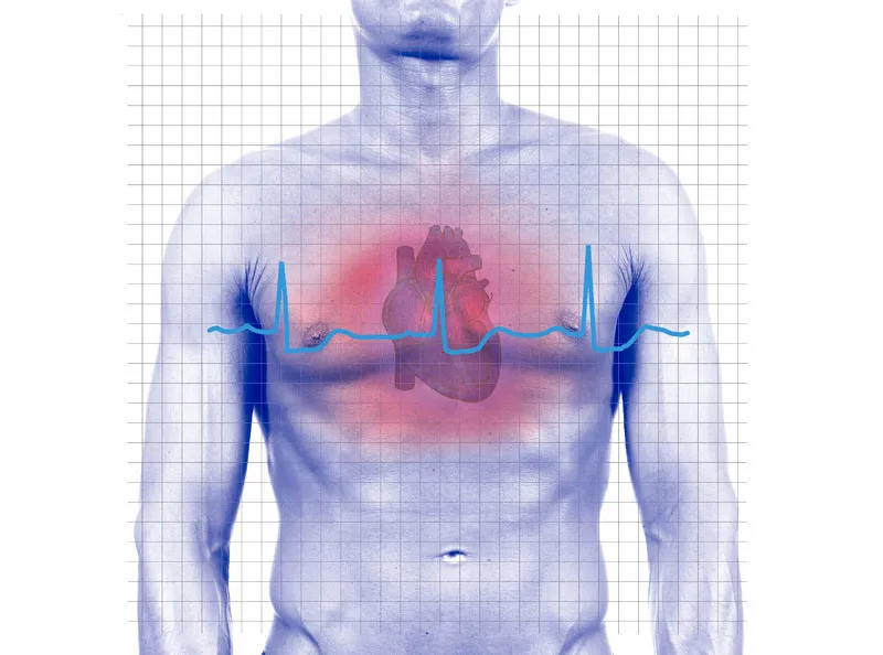
Today’s technology for firing up the heart has been extremely useful, but has some limitations, Jenkins said.
For a typical pacemaker, electrodes – strips of metal – are inserted into heart tissue and connected via wire to a box, which is implanted under a person’s collarbone. (The box contains a computer chip to monitor the heart’s electrical activity and power to deliver electric pulses when needed to stimulate the heart.)
One issue with this setup: The metal strips are stuck directly into the heart, which could damage tissue. This could be particularly important for infants and children.
In addition, electricity-based pacemakers can’t be used during surgery when magnetic resonance image (MRI) is being used – a combination that is becoming more frequent today as doctors watch the images while performing robotic surgery, Jenkins said.
Light pacing
“I don’t see optical pacing completely taking over for electrical pacing, but I think in applications where optical pacing may have an advantage it might be adopted,” Jenkins said.
The device may prove beneficial in lab work to better understand how a heart develops and ultimately determine the cause of congenital heart failure. The advantage of optical pacing here is its precision, which would give a clear picture of cause and effect.
“If I send electrical current into tissue it kind of goes anywhere and I excite a huge region; whereas if I focus light I could theoretically just excite a single cell,” Jenkins said.
“Another advantage of optical pacing is that it’s compatible with MRI imaging,” Jenkins added.
And because the new setup is non-invasive, it would lend itself to more sensitive procedures. “I don’t have to stick a lead into the tissue and perhaps damage some cells,” Jenkins said. Instead, the fiber-optic could be fed through a catheter up to the heart.
Jenkins and his colleagues detailed their research online Aug. 15 in the journal Nature Photonics.