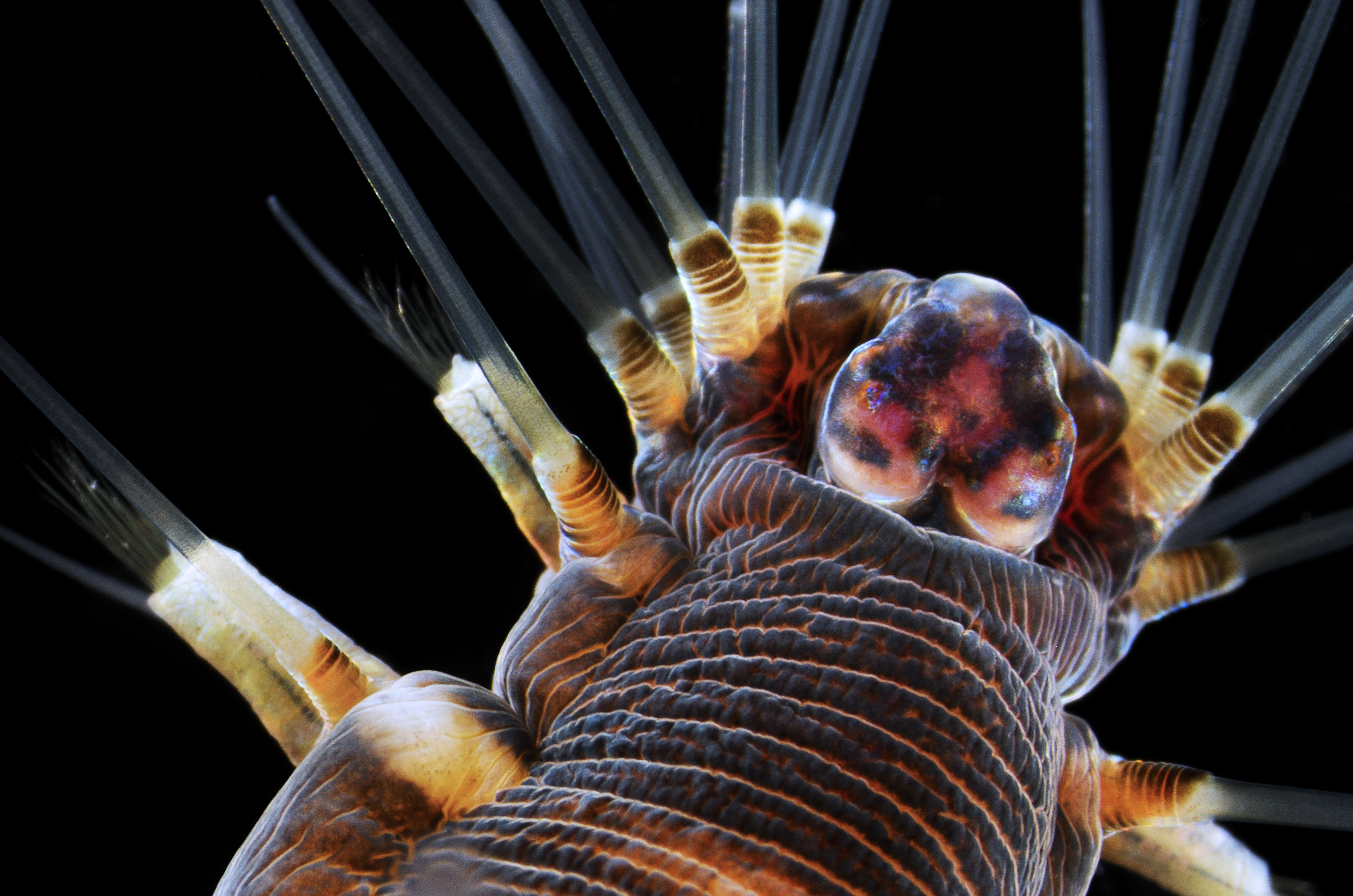
This amazing image of a marine worm took third place at Nikon's 2013 Small World Photomicrography competition.
Dr. Alvaro Esteves Migotto from Universidade de São Paulo, Centro de Biologia Marinha in São Paulo, Brazil, submitted the striking photo. To create this image of a marine worm, Migotto used dark-field microscopy, in which a dark field microscope blocks off the light source, causing light to scatter as it hits the specimen. This technique is ideal for making objects with refractive values (how light travels through a medium) similar to the background appear bright, including unstained or transparent organisms.
Because the animal was alive and active during its "photo shoot," Migotto lit it with two flashes, so the motion of the specimen would not blur the image.
Follow LiveScience @livescience, Facebook & Google+.
Get the world’s most fascinating discoveries delivered straight to your inbox.


