Spectacular Brain Images Reveal Surprisingly Simple Structure

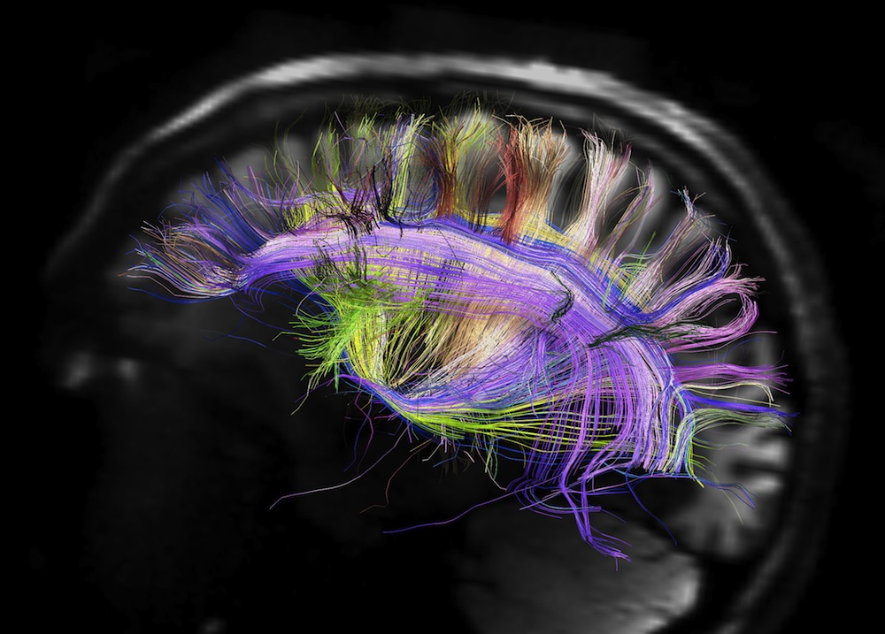
Stunning new visuals of the brain reveal a deceptively simple pattern of organization in the wiring of this complex organ.
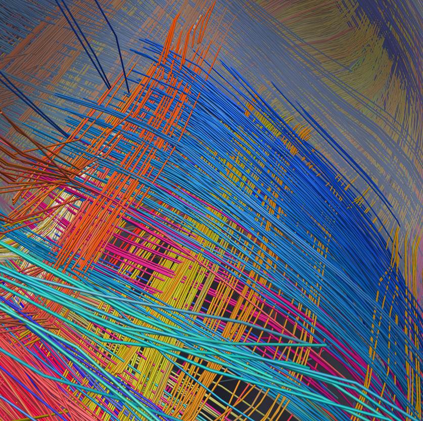
Instead of nerve fibers travelling willy-nilly through the brain like spaghetti, as some imaging has suggested, the new portraits reveal two-dimensional sheets of parallel fibers crisscrossing other sheets at right angles in a gridlike structure that folds and contorts with the convolutions of the brain.
This same pattern appeared in the brains of humans, rhesus monkeys, owl monkeys, marmosets and galagos, researchers report today (March 29) in the journal Science.
"The upshot is the fibers of the brain form a 3D grid and are organized in this exceptionally simple way," study leader Van Wedeen, a neuroscientist at Harvard Medical School and Massachusetts General Hospital, told LiveScience. "This motif of crossing in three axes is the basic motif of brain tissue." [Inside the Brain: A Journey Through Time]
The organized brain
The surface of the brain contains about 40 billion nerve cells, each making about 1,000 connections in a pattern that brain researchers have yet to decipher, said Marsel Mesulam, the director of the Cognitive Neurology and Alzheimer's Disease Center at Northwestern University. Mesulam, who was not involved in the study, called Wedeen's work "very exciting."
"There can be no more fundamental question in philosophy, in psychology," Mesulam told LiveScience. "The human brain is the single most complex device in the known universe, and it works by nerve cells talking to each other. If we can't figure out how they decide who to talk to and what they tell each other, we just don't understand how the brain functions."
Get the world’s most fascinating discoveries delivered straight to your inbox.
Using a technique he developed called diffusion spectrum magnetic resonance imaging (MRI), Wedeen traced the movement of water molecules along the intersections of brain fibers (the cellular projections that form the brain's communication network), tracking the orientation of each fiber at each crossing.
"What emerged was astonishing," Wedeen said. "What emerged was that the set of fibers that crossed a given fiber, invariably — and that's a really strong invariably — look like mutually parallel fibers all coming in like the teeth of a comb and crossing it in one direction." [See video of the brain structure]
Animal studies had suggested this pattern might exist, and researchers already knew that the nerve cells in the spinal cord and brain stem were organized in very structured parallels and perpendiculars even in humans (consider the long nerve fibers that run down the backbone and then branch out perpendicularly from the vertebrae). But it's difficult to get high-resolution scans of fiber connectivity in the human cortex, given that humans tend to become uncomfortable if left in an MRI scanner for more than 45 minutes or so, Wedeen said. For that reason, images of human brain connections have tended to look like tangled spaghetti, he said.
Wedeen and his colleagues scanned four types of primate brains from deceased animals, enabling them to image the brains for up to 48 hours, as well as brains from living human subjects using a new scanner that can achieve 10 times the resolution of conventional MRI machines. Using special software, the researchers then reconstructed three-dimensional images of the brain-fiber pathways.
"Looking across multiple species, it emerged that the pattern was substantially similar," Wedeen said. "When you went from primates with small brains to primates with big brains … the rules were the same, but they were being applied more diversely and with more layers in the larger, more complex brains."
Adaptable brain
The finding of clear up-down, front-back and side-to-side organization in the brain makes sense, Wedeen said, given that the brain has had to rewire both evolutionarily (to form the specialized brains humans boast today) and during its lifetime (as it grows and learns, for example). If the organization of communication were chaotic, that wouldn't work.
"It's like rewiring your basement at random," Wedeen said. "First thing that happens, house burns down, you die."
In other words, adapting a complexly wired brain that will still allow the next generation to survive would be next to impossible.
"If you try to picture what would happen if you tried to turn one spaghetti brain into a different spaghetti brain, you realize you would need an impossibly knowledgably intelligent designer standing above the brain and rewiring it," Wedeen said.
With an organized grid structure, however, evolution can easily build on what came before — adding in a more complex forebrain in humans versus our monkey relatives, for example.
More work should be done to link the imaging methods of Wedeen with traditional neuroanatomy methods to confirm the findings, Mesulam said. Wedeen plans to expand the map of the human brain into more detail. It's also important to understand the relationship between a brain's structure and its function, he said. Understanding the structure of a typical brain would ultimately help scientists comprehend what happens when brain development goes wrong, as in Alzheimer's or mental illness.
"Say somebody comes to you with their 2-year-old and they say, 'My 2-year-old is just not looking me in the eyes'. Is this the first sign of Asperger's or just an individual difference?" Wedeen said. "You'd know how to begin. You'd know what you were doing."
You can follow LiveScience senior writer Stephanie Pappas on Twitter @sipappas. Follow LiveScience for the latest in science news and discoveries on Twitter @livescience and on Facebook.

Stephanie Pappas is a contributing writer for Live Science, covering topics ranging from geoscience to archaeology to the human brain and behavior. She was previously a senior writer for Live Science but is now a freelancer based in Denver, Colorado, and regularly contributes to Scientific American and The Monitor, the monthly magazine of the American Psychological Association. Stephanie received a bachelor's degree in psychology from the University of South Carolina and a graduate certificate in science communication from the University of California, Santa Cruz.
 Live Science Plus


