
 Live Science Plus


Inside the Brain: A Photo Journey Through Time
The Human Brain

The brain has long boggled the mind with its complexity, which is probably best summed up by Carl Sagan in "The Cosmos," when he said, "The brain is a very big place in a very small space." With modern technology, scientists are peering deeper and closer than ever before at the tangle of neurons and their billions of connections. Here's a peek at what the brain looks like, from antiquity to present-day.
Portraits of the Mind

In the book, "Portraits of the Mind: Visualizing the Brain from Antiquity to the 21st Century" (Abrams 2010), astonishing images that reveal both the complexity and beauty of the brain. And through time as brain-imaging technology comes online, scientists have new ways of seeing and interpreting the brain. Check out some of the amazing photos from the book.
Canine Scents

This 1875 drawing showing a dog's olfactory bulb was completed using a staining method named after Camillo Golgi in which certain chemicals are injected into nervous tissue so they can be seen. Some say its application to the study of brain tissue represents the beginning of modern neuroscience.

Dripping Dendrites

While all cells in the body hold the same genome, only a particular set of its genes get turned on in various cells; each type of neuron switches on a gene set that defines its character.
In this picture, a gene called JAM-B had been switched on, which then turned on a fluorescent protein to reveal a small group of brain cells. The resulting image shows that all of the neurons' projections called dendrites are aligned in the same direction; moreover, these retinal neurons are known to detect only objects moving in an upward direction.
Baroque Blood Vessels

A scanning electron microscope (SEM) image zooms in on the baroque branching structures that send blood to the human brain's cortex. The vessels are organized such that the large blood vessels surround the surface of the brain (top of image), sending thin, dense projections down into the depths of the cortex (bottom of image).
View of a Stroke
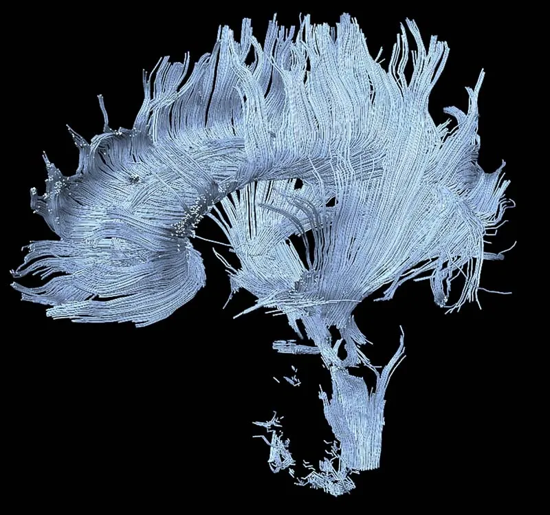
A brain-imaging method called diffusion MRI (magnetic resonance imaging) is relatively new to the field of neuroscience, though it shows promise as a diagnostic tool. Here, an image taken from the brain of a patient who suffered a stroke in the thalamus and midbrain, resulting in damage to certain axons (some are visible at the bottom of the image).
Get the world’s most fascinating discoveries delivered straight to your inbox.
Mouse Brain

A cross-section of a mouse's hippocampus — one of the brain's memory centers — reveals its intricate network of neurons, whose soma are shown as small circles. The hippocampus is seen here nestled directly beneath the neocortex, which is the outer layer of the cerebral hemispheres.
Spiny Neuron
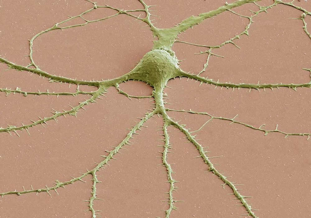
Most neurons have three parts: an axon, a cell body called a soma and dendrites. This scanning electron microscope (SEM) image shows a soma with dendrites (and their spines) radiating from it. To create SEM images, a beam of electrons is scanned across the surface of a sample, and a detector keeps track of electrons bouncing off its surface to reveal the specimen's outer shape.
Artsy Brain Cells

Here, two types of cells in the cerebellum are shown: glia and Purkinje neurons. The cells can be distinguished because of a method that relies on the body's immune system and its antibodies — proteins that recognize and latch onto "foreign substances." Biologists now use antibodies to reveal where certain proteins are found in the brain. Here, red is an antibody staining of a protein that's found in glia cells, while green reveals a protein called IP3, of which Purkinje neurons are chockfull.
Color My Cerebellum

The colored splotches reveal so-called presynaptic terminals, or junctions through which neuron signals are sent, formed by the cerebellum's axons.
Brainbow

While Golgi's staining method did wonders for finding structures hidden in a tangle of neurons, it couldn't distinguish individual brain cells that were illuminated in the same color.
Enter a bit of genetic trickery called Brainbow: Robert Tsien and other chemists tinkered with and discovered fluorescent proteins responsible for the different colors emitted by various sea creatures (such as corals and jellyfish). By coaxing different sets of neurons or even different individuals of a species (say a male and female) to express different proteins, scientists could pick out the cells by the color they glowed.
Here, several motor-neuron axons (slender projections on neurons that transmit signals to other neurons) travel side by side as they lead to the muscles whose contractions they regulate.
Jeanna Bryner is managing editor of Scientific American. Previously she was editor in chief of Live Science and, prior to that, an editor at Scholastic's Science World magazine. Bryner has an English degree from Salisbury University, a master's degree in biogeochemistry and environmental sciences from the University of Maryland and a graduate science journalism degree from New York University. She has worked as a biologist in Florida, where she monitored wetlands and did field surveys for endangered species, including the gorgeous Florida Scrub Jay. She also received an ocean sciences journalism fellowship from the Woods Hole Oceanographic Institution. She is a firm believer that science is for everyone and that just about everything can be viewed through the lens of science.