It's quick and easy to access Live Science Plus, simply enter your email below. We'll send you a confirmation and sign you up for our daily newsletter, keeping you up to date with the latest science news.
A choice between drinking a sugary beverage and getting radiation is a no-brainer for anyone, especially cancer patients, who need to get their bodies scanned regularly to check for tumors. New research suggests that a high dose of sugar makes cancerous tumors stand out on magnetic resonance imaging (MRI) scans, meaning such scans could sometimes be used instead of conventional positron emission tomography (PET) scans, which require radioactive material.
The researchers tested this MRI method by scanning human tumors implanted in mice, and the results were as good as those produced by PET scan using radioactive material.
"The method could offer a cheap, safe alternative to existing methods for detecting tumors," said study researcher Simon Walker-Samuel, of the University College London. The radioactive material used in PET scans is expensive to manufacture, and pose a risk, especially to vulnerable groups such as children and pregnant women.
The new method takes advantage of the fact that uncontrollably dividing cancer cells consume far more sugar than normal cells, making them stand out in an MRI scan that is aimed at detecting glucose, a type of sugar.
The amount of sugar a patient needs to consume before undergoing such a scan would be about the amount found in half of a chocolate bar, the researchers wrote in their study, which was published July 7 in the journal Nature.
The researchers are now using the new method on cancer patients, to take the first experimental steps toward running a clinical trial in the future.
"It's looking promising," Walker-Samuel said. "The patients lie in the scanner, and drink a sugary drink. And then, up to an hour later, we start seeing the accumulation of glucose in tumors."
Sign up for the Live Science daily newsletter now
Get the world’s most fascinating discoveries delivered straight to your inbox.
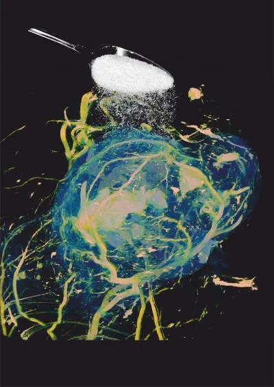
Bright yellow regions show increased sugar uptake at the edges of the tumor compared with blue central regions.
(Image credit: UCL)
However, there are hurdles to using the scans in patients. For example, the MRI machines used on patients are as much as three times weaker than those used in labs for studies, like the one the researchers used on mice. The weaker machines may not detect as clear and strong of a signal from glucose, the researchers said, so they are experimenting with ways work around this.
An MRI machine works by inducing a strong magnetic field, which excites the protons in water molecules within the body. The machine then sends another wave of energy, and measures how long it takes for the protons to return to their normal state. A computer uses this data to construct an image of the body tissue.
In the new method, the researchers used a wave with a frequency slightly different from what they use to measure water, which makes the scan able to detect where the most glucose is concentrated.
For any MRI, "the sensitivity and specificity isn't always as good as it can be, and we're always looking for ways of improving that," Walker-Samuel said.
It's difficult to say how long it might take before the new method could be used in patients, the researchers said, but they are hopeful that it will be done within three to five years.
"It's usually straightforward to translate [new imaging techniques] from a preclinical animal setting into a clinical setting," Walker-Samuel said, noting that although ethical considerations and licensing often slow down the process, the prospects look better in this case. "That's not such an issue, I think, when we are doing something like just asking the patient to drink a sugary drink," he said.
Bahar Gholipour is a staff reporter for Live Science covering neuroscience, odd medical cases and all things health. She holds a Master of Science degree in neuroscience from the École Normale Supérieure (ENS) in Paris, and has done graduate-level work in science journalism at the State University of New York at Stony Brook. She has worked as a research assistant at the Laboratoire de Neurosciences Cognitives at ENS.