
 Live Science Plus


12 Amazing Images in Medicine

Introduction

Modern medicine has accomplished some incredible feats, including helping people to survive injuries and diseases that might have seemed impossible to survive a few decades ago.
Doctors and researchers have captured images of some of these cases, and we've rounded them up, along with other images of unusual symptoms and features of conditions.
Here's a look at five amazing images in medicine.

A fence pole through the head

This X-ray shows 28-year-old Las Vegas man Andrew Linn, who survived a 2010 car accident that pushed a 2-inch-thick metal fence pole through his mouth and out through the back of his neck, according to a report of his case in the Las Vegas Review-Journal.
Linn "was very calm and didn't appear in any pain," surgeon Dr. Jay Coates, of University Medical Center, told the Review-Journal. The medical team at was able to remove the pole without damaging either of two major blood vessels — the carotid artery and jugular vein — in the neck region.
Star shaped cataract

A man in Austria developed a cataract shaped like a star in his eye after he was punched. The man went to his doctor because his vision had worsened over the previous six months, according to doctors who treated the man. The patient said he'd been punched nine months earlier, the doctors wrote in their report. It's very common for cataracts to form after the eye takes a hit, said Dr. Mark Fromer, an ophthalmologist at Lenox Hill Hospital in New York City, and eye surgeon for the New York Rangers hockey team. Punches and the balls used in sports are most often the cause, but bumps from air bags and steering wheels have also created cataracts, Fromer said. When the eyeball is struck, the energy of the blow sends shock waves through the eye that can disrupt the nature of the eye's lens, causing it to become opaque in regions, he explained. In most cases, cataracts look more like a vaguely shaped cloud, and can be white or yellowish. The case is reported in April 2013 in the New England Journal of Medicine.
Gold needles in the knee
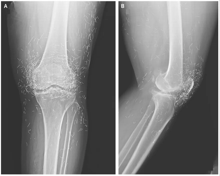
When doctors examined an X-ray image of the knees of a woman experiencing severe joint pain, they found a gold mine: hundreds of tiny gold acupuncture needles left in her tissue.
Get the world’s most fascinating discoveries delivered straight to your inbox.
The 65-year-old South Korean woman had previously been diagnosed with osteoarthritis, a condition in which the cartilage and bones within the joints degrade, causing pain and stiffness.
But when pain relievers and anti-inflammatory drugs didn't alleviate the pain in her knees and only caused stomach discomfort, she had turned to acupuncture, she told her doctors. The needles, which were presumably made of gold, were intentionally left in her tissue for continued stimulation, her doctors wrote in the New England Journal of Medicine in December 2013.
A hairy eyeball

This picture shows a mass called a "limbal dermoid" on the right eye of a 19-year-old man. Limbal dermoids are solid masses and frequently contain hair shafts.
The man had come to a clinic because he had discomfort when he blinked, and felt as though there was something in his eye, according to Dr. Ali Mahdavi Fard and Dr. Leili Pourafkari, of the Tabriz University of Medical Sciences in Iran, who reported his case in the New England Journal of Medicine in January.
He'd had the mass since birth, but it had gradually grown. In his left eye, he had 20/20 vision, but in the right eye, his vision was 20/60, according to the case report.
The man had the mass removed in surgery for cosmetic reasons, but his vision did not improve.
An accidental swallowing of a knife

This X-ray shows a knife that became stuck in a 30-year-old woman's esophagus and stomach, causing her "chest discomfort," according to a report of her case.
The woman had previously had the eating disorder bulimia, and had inserted the knife into the back of her mouth in order to demonstrate that she had lost her gag reflex. But she laughed unexpectedly, and the knife fell into her body, according to Dr. Aida Venado and Dr. Sarah Prebil, of Emory University School of Medicine, who reported the woman's case in the New England Journal of Medicine in 2012.
Doctors were able to remove the knife in a procedure called an esophagogastroduodenoscopy, in which a small camera is used to view the esophagus. The woman recovered, and she was able to eat without complications, according to the report.
"Half-half" blisters

This image of unusual blisters on a 66-year-old man was captured at Leeds General Infirmary in England.
The blisters contained both clear pus and yellow fluid, and were found on the man's trunk, according to Dr.Michael O'Connell and Dr. Victoria Goulden, who reported the man's case in the New England Journal of Medicine in 2012.
After a biopsy, the man was diagnosed with a condition called subcorneal pustular dermatosis. A similar condition can be caused by drugs, but trigger in this man's case was unclear, according to the case report.
The man was treated with steroid medications and recovered.
Beer bottle in the colon

This X-ray was taken of an intoxicated 35-year-old man who went to an emergency room in Mexico with rectal bleeding and abdominal pain.
Doctors examined his abdomen and found a mass, but no signs of trauma. A rectal exam revealed a foreign body that could not be seen, so once the patient was well enough, he was sent to be X-rayed, according to Dr. Roberto Flores-Suarez and Dr.Jorge Reyes-del Valle, of the State Health Institute of Mexico, who reported the man's case in the New England Journal of Medicine in 2010.
The bottle was removed in surgery, and the man was treated with antibiotics and pain relievers. He eventually recovered.
Skin cancer in the gums
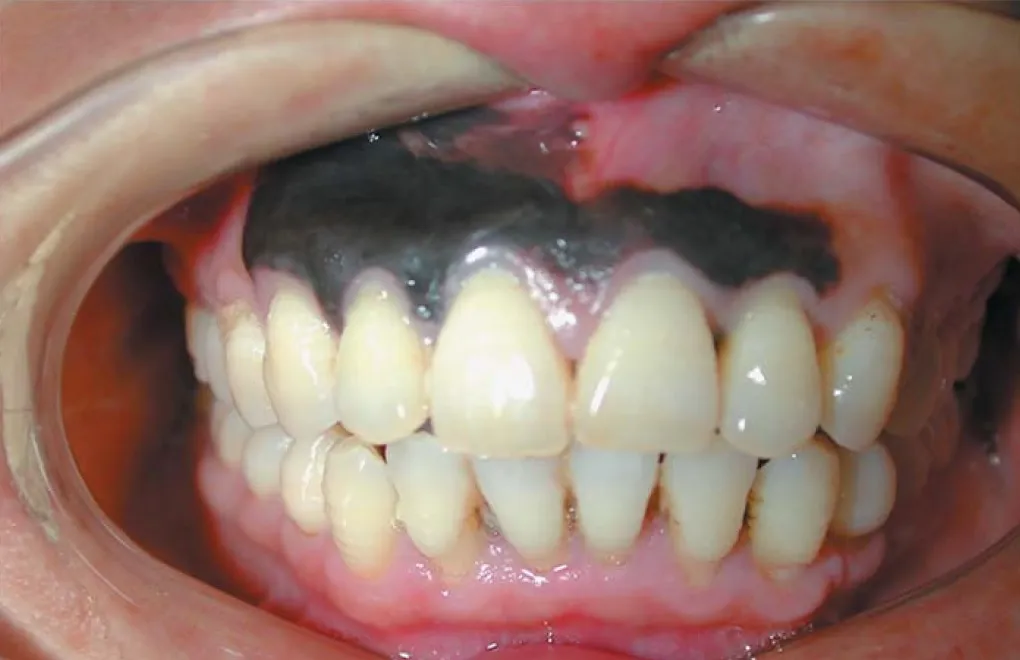
A 45-year-old man in China developed a large, dark discoloration of his upper gums that doctors diagnosed to be a rare type of melanoma, a cancer that usually occurs in the skin.
This type of melanoma, medically known as mucosal melanoma, constitutes less than 1 percent of all melanoma cases, Dr. Susan Muller, a professor of pathology at Emory University who wasn't involved with the case, told LiveScience.
Pigmented dark spots in the mouth are not uncommon, but are usually benign (non-cancerous). In this patient, however, the large area of discoloration alarmed the doctors, and they performed a biopsy.
The biopsy showed the dark lesion was, indeed, cancerous, and doctors removed the gums and parts of the man's upper jaw. A report of the case was published in the Oct. 9 issue of The New England Journal of Medicine.
Pellets in the appendix
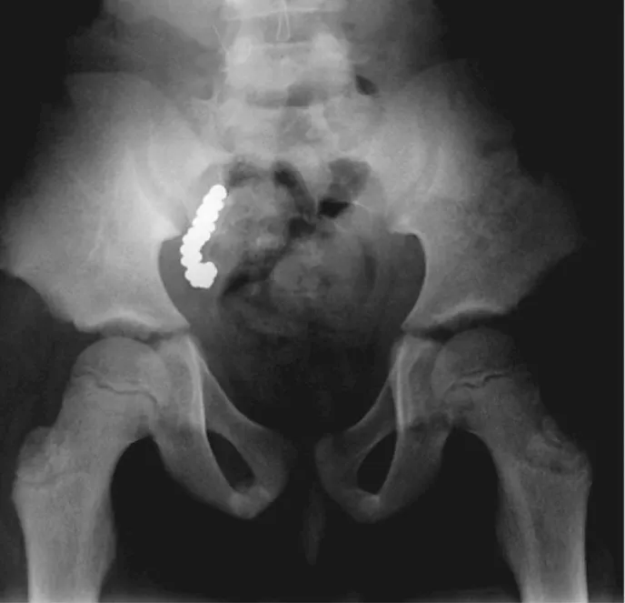
An 8-year-old boy in Australia had high levels of lead, a toxic metal, in his blood for more than two years. No one knew why, until doctors found 57 lead pellets trapped in his appendix.
The boy had been showing unusually hyperactive behavior for months, which prompted the doctors to check for lead poisoning. The tests revealed the boy had about five times the highest lead levels normally found in humans. But the source of the poisonous chemical remained unknown until the boy developed a stomachache and doctors X-rayed his abdomen.
"It's one of those things you only see once in a lifetime," Dr. Ibrahim Zardawi, the pathologist who examined the appendix, told LiveScience. "I've been in medicine for almost 40 years now, and had never seen anything like this."
Later, the boy and his siblings told the doctors that they had been eating the pellets they found in the geese that their family regularly hunted.
The case was reported in the Aug. 8 issue of the New England Journal of Medicine.
Inhaled dart
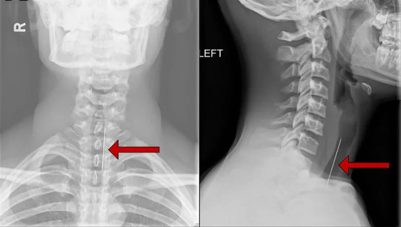
A 15-year-old boy in Ohio accidentally inhaled a dart from a blowgun, which made him cough for three hours before sending him to the emergency room, according to a report of his case.
The dart got trapped in the boy's airway when he inhaled while holding the blowgun in his mouth. A blowgun, essentially a narrow tube, is designed so that people can propel darts outward by the force of an exhaled breath.
At the emergency room, X-rays of the boy's airway revealed the dart. After further questioning, the boy admitted to using the blowgun.
The doctors inserted a tube down the boy's throat to get a better view of the object in his airway, and removed the dart. The boy left the hospital without any serious complications, but inhaling objects can be life-threatening if it blocks the airway. Sharp objects can also puncture the airway or damage the voice box, the doctors told LiveScience.
The report is published in the July 22 issue of the journal Pediatrics.
