


Tracking Cellular Sugar Traffic
This Research in Action article was provided to LiveScience in partnership with the National Science Foundation.
A glow-in-the-dark sugar coating sounds like the feature of an awesome pastry. Turns out it also has a place in zebrafish embryos, whose sugary cell surfaces scientists have tagged with fluorescent proteins to track cell traffic.
Virtually all cells in a zebrafish — and a human — have a coating called a glycocalyx. It's made of complex sugars called glycans, which are involved in important processes like cell communication, immune response and early development. Because of their roles, a growing number of researchers are turning to glycans to understand and treat diseases.
A team of researchers led by chemical biologist Carolyn Bertozzi at the University of California, Berkeley, tagged these glycans with fluorescent molecules to get a closer look at how zebrafish embryos develop. The team used zebrafish because their embryos are transparent, and as a common model organism, zebrafish can tell us something about human development. The imaging technique is non-toxic, meaning it can be used on living cells.
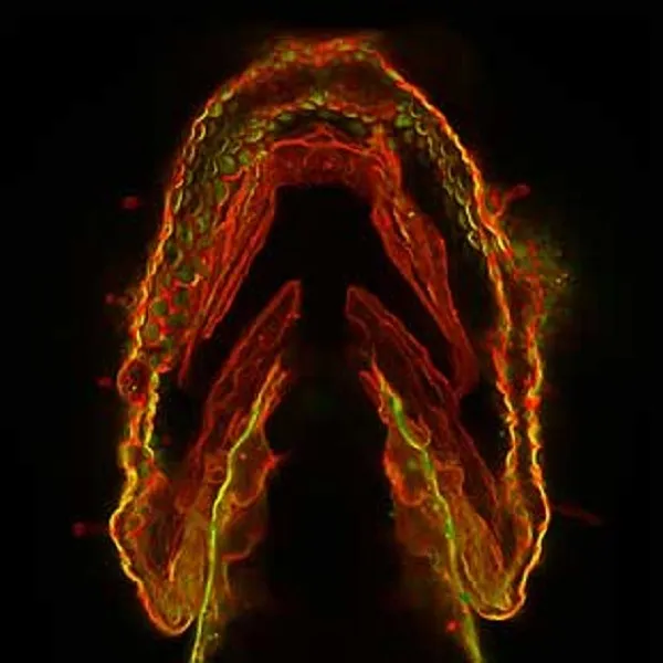
In the picture above, the jaw of a three-day-old zebrafish embryo glows red where recently produced glycans surround cell surfaces. Green-glowing glycans produced earlier in development have already migrated into the cells. Where old and new glycans congregate, the jaw shines yellow.
Investigating glycan traffic patterns is helping scientists understand how tissues develop in zebrafish, which could extend to other vertebrates. This work could uncover early markers for disease; for example, scientists suspect that glycan patterns are different in cancer cells than they are in normal cells. Glycans also play a role in tuberculosis, HIV, muscular dystrophy, malaria and other diseases. The influenza virus — like virtually all viruses and bacteria — attacks by attaching to sugars on the host cell's surface.
This research was supported by the National Institutes of Health. To see more cool images and videos of basic biomedical research in action, visit http://publications.nigms.nih.gov/biobeat/gallery/index.html.
Get the world’s most fascinating discoveries delivered straight to your inbox.