
 Live Science Plus


Bionic Eyes Plug Directly into the Brain

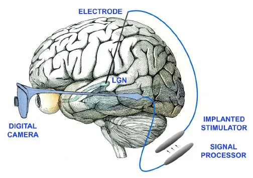
Researchers have set their sights on bypassing the normal routes of bionics to hook video cameras deep into the brain, allowing the blind to see. The bionic eye system has already proved promising in monkeys. The goal is to one day provide vision for blind people using twin video cameras worn as a pair of glasses that transmit signals wirelessly to an implant in the brain. Decades of studies have tried to develop prostheses that restore sight. One approach generates images by electrically stimulating healthy neurons in the retina, the light-sensitive tissue lining the inner eyeball , and thus mimic the effects of inbound light. The other aims to electrically stimulate cells in the cortex, the outer layer of the brain where visual signals are processed. Both approaches have drawbacks and have achieved limited success. The retina is a very delicate, fragile membrane that is prone to damage. And the complexity within the cortex makes it more difficult to generate intelligible pictures with the second method. With retinal stimulation, each neuron that gets stimulated is responsible for a single dot in any image that is seen. In cortical stimulation, "you stimulate one site in the cortex, you get one spot of light, you stimulate another site you get another spot, but if you stimulate both at once you don't necessarily get the sum of the two, but you might get a third spot," explained computational neuroscientist computational neuroscientist Nicholas Hatsopoulos of the University of Chicago in Illinois. "You can't really connect the dots." Going deeper So neuroscientists John Pezaris and R. Clay Reid at Harvard Medical School concentrated on a region deep in the brain called the thalamus, which relays sensory data between the cortex and the rest of the body. They focused on a part of the thalamus known as the lateral geniculate nucleus, which relays visual signals. The researchers examined the eye movements of rhesus monkeys with normal vision in response first to points of light on a computer screen and then to electrical stimulation of the thalamus. During electrical stimulation, the monkeys shifted their gazes to points on the screen corresponding to the location of electrodes in the brain. This suggests the animals treated the electrical stimuli as normal visual images, findings detailed online this week in the Proceedings of the National Academy of Sciences. The thalamus is easier to stimulate than the retina and much less fragile, the researchers said. At the same time, the targeted neurons should behave more like those in the retina than those in the cortex, making it simpler to generate intelligible images. "Their approach offers the best of both worlds, the benefits of the other two methods without their drawbacks," said Hatsopoulos, who did not participate in this research. Challenges Pezaris did note the part of the thalamus they focused on is often thought of as difficult to access surgically, since it is deep in the brain. However, "implanting an electrode into the deeper parts of the brain has become a routine procedure," Pezaris told LiveScience. "These techniques are used in the clinic to treat things like Parkinson's disease, and are being actively explored for treating major depression, obsessive compulsive disorder and other conditions. While any brain surgery has some risk, it seems that neurosurgeons have overcome the hurdles so that it can be performed to acceptable standards of safety." For these initial experiments, the researchers only used two electrodes, each just 35 microns across or roughly a third the width of a human hair, to generate two spots the monkeys could see. Pezaris said their immediate goal is to use more electrodes to create more complex images. The lateral geniculate nucleus is a relatively tiny structure that might not tolerate having thousands of electrodes implanted into it for a high-resolution picture, but he noted advances in electronics might lead to even finer wires to surmount this challenge.
- Blind Cats Get Implants
- Machine Offers Sight to Some Blind People
- Visual Response Restored in Blind Mice
Get the world’s most fascinating discoveries delivered straight to your inbox.
