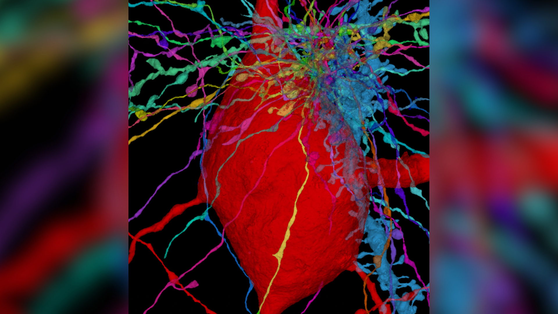
3D map plots human brain-cell 'antennae' in exquisite detail
A new map of 56,000 cells in the outer layer of the human brain could inform research into a whole class of diseases.

Get the world’s most fascinating discoveries delivered straight to your inbox.
You are now subscribed
Your newsletter sign-up was successful
Want to add more newsletters?

Delivered Daily
Daily Newsletter
Sign up for the latest discoveries, groundbreaking research and fascinating breakthroughs that impact you and the wider world direct to your inbox.

Once a week
Life's Little Mysteries
Feed your curiosity with an exclusive mystery every week, solved with science and delivered direct to your inbox before it's seen anywhere else.

Once a week
How It Works
Sign up to our free science & technology newsletter for your weekly fix of fascinating articles, quick quizzes, amazing images, and more

Delivered daily
Space.com Newsletter
Breaking space news, the latest updates on rocket launches, skywatching events and more!

Once a month
Watch This Space
Sign up to our monthly entertainment newsletter to keep up with all our coverage of the latest sci-fi and space movies, tv shows, games and books.

Once a week
Night Sky This Week
Discover this week's must-see night sky events, moon phases, and stunning astrophotos. Sign up for our skywatching newsletter and explore the universe with us!
Join the club
Get full access to premium articles, exclusive features and a growing list of member rewards.
Tiny, hairlike "antennae" protrude from the surface of brain cells, and now, scientists have unveiled a detailed map of these wires across the whole human cortex. They hope the new map will guide future research into a class of diseases that cause these structures to malfunction.
The hairlike structures, known as cilia, are actually found on the surface of most eukaryotic cells, meaning complex cells that house their DNA in a nucleus. Some cilia can move; for example, cilia in the lungs collectively beat to clear the airways of harmful pathogens. Others, called primary cilia, are immobile and instead act like antennae, sensing signals from their environment and passing them to the nucleus of the cell.
In the brain, neurons and their associated helper cells, called glia, need primary cilia to effectively receive and send signals. However, until now, little has been known about cilia's structure or organization in the brain or how they slot into the network of interactions between neurons in the brain known as the "connectome."
In a new study, published Oct. 27 in the journal Neuron, scientists constructed a three-dimensional map of the primary cilia in the brain's outer layer, or cortex, which is part of the cerebrum, the largest portion of the brain. The researchers hope the new map, which details 56,000 cells, will guide research into ciliopathies, a class of diseases tied to disruptions in cilia function.
Related: Most detailed human brain map ever contains 3,300 cell types
"There are a number of cognitive difficulties associated with mutations in the proteins that make up cilia," co-senior study author, Dr. Jeff Lichtman, a professor of molecular and cellular biology at Harvard University, told Live Science in an email. "These studies may suggest that who a cilia contacts may be important in their normal function and when that function goes awry in disease."
To create the new map, the researchers pieced together images of the six layers of the human cerebral cortex; the images depicted "slices" of a tissue sample that had been collected during a surgery to treat an adult patient with epilepsy. One by one, the team looked at the cilia that emanated from each brain cell and studied how they interacted with one another.
Get the world’s most fascinating discoveries delivered straight to your inbox.
The cilia differed in size and shape depending on the type of cell they interacted with and the layer of the cortex where they were located. Cilia were also a key component of the structure of some synapses — the junctions between neurons that allows these cells to communicate with each other — suggesting that cilia are firmly embedded in the brain's connectome.
The diversity of the cilia raises the "intriguing possibility" that different types of cilia in the cortex can regulate the activity of neurons in unique ways, the authors said.
Going forward, the team would like to delve further into how these primary cilia influence neural circuits in the brain and how this knowledge could be used to treat neurological disorders where cilial function is impeded, co-senior study author E. S. Anton, a professor of neuroscience at the University of North Carolina School of Medicine, told Live Science in an email.
Ever wonder why some people build muscle more easily than others or why freckles come out in the sun? Send us your questions about how the human body works to community@livescience.com with the subject line "Health Desk Q," and you may see your question answered on the website!

Emily is a health news writer based in London, United Kingdom. She holds a bachelor's degree in biology from Durham University and a master's degree in clinical and therapeutic neuroscience from Oxford University. She has worked in science communication, medical writing and as a local news reporter while undertaking NCTJ journalism training with News Associates. In 2018, she was named one of MHP Communications' 30 journalists to watch under 30.
 Live Science Plus


