Brain scans of babies in the womb may reveal whether a child is at risk for developing autism later in life, early research suggests.
A small study of 39 fetuses found that, by 25 weeks of gestation, certain brain regions looked different in the unborn babies who went on to be diagnosed with autism compared with those who were not diagnosed with the condition.
Specifically, prenatal MRI scans showed that the insular lobe — which may play a role in perceptual awareness, social behavior and decision-making — was larger in volume in the babies who would later be diagnosed with autism, compared with the insular lobes of children who were not diagnosed with autism.
Latest Videos From
Children in the autism group also had larger volume in a brain region called the amygdala in prenatal scans — a finding that jibes with earlier results showing larger amygdalae in toddlers with autism.
"These results make it clear that we need to focus on these promising regions as potential biomarkers and find out the reason for these alterations," study first author Alpen Ortug, a postdoctoral research fellow at Massachusetts General Hospital, Harvard Medical School, told Live Science in an email.
Related: What is the amygdala?
The findings add to a growing body of evidence that the disease processes involved in autism may begin early in development, the researchers said.
Still, much more research is needed to confirm the findings, which were presented Tuesday (April 5) at the Experimental Biology (EB) 2022 meeting in Philadelphia. The study has not yet been published in a peer-reviewed journal.
Autism spectrum disorder (ASD) is a developmental disorder that affects how a person communicates, interacts socially, learns and behaves, according to the National Institutes of Health (NIH).
Early detection and treatment of autism can greatly improve outcomes for patients, according to the NIH. But currently, the earliest that autism can be reliably diagnosed is about 18 months of age, the researchers said.
Previous studies have found brain differences in infants that go on to develop autism. For example, a study published March 25 in The American Journal of Psychiatry found that the amygdala may grow too fast in babies between 6 and 12 months of age prior to their diagnosis of autism, Live Science previously reported.
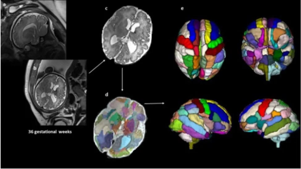
In the new study, the researchers examined whether prenatal brain scans could help spot potential markers of autism even before birth. They analyzed 39 fetal MRI brain scans, which were performed at Boston Children's Hospital. The MRI scans were originally conducted because the fetuses were suspected to have a developmental condition based on ultrasound results, but the ultrasounds were not sufficient to confirm the diagnosis, Ortug said.
Among these patients, nine children were later diagnosed with autism, and 20 children had typical development. Ten of the children did not have autism but had other health conditions, such as developmental disorders affecting the cardiovascular system. The MRI scans were analyzed retrospectively, meaning after the children's diagnoses.
The researchers used a computer programming method to segment the brain scans into different regions and then compared the segmented regions among the different groups.
They found the biggest differences in the insular lobe, with significantly larger volumes in the autism group compared with the other groups. This finding agrees with previous studies that have found changes in the insular lobe in adults with autism, and suggests these changes may start in the womb, the researchers said.
Dr. L. Eugene Arnold, a professor emeritus of psychiatry and behavioral health at The Ohio State University Wexner Medical Center who was not involved with the new study, told Live Science that the new study was small and that the findings need replication but that the results are in line with other reports of various prenatal differences linked with autism. For example, a study published in January in the journal Brain that examined prenatal ultrasounds found that children who went on to develop autism were much more likely to have anomalies in their heart, kidneys and head seen on the ultrasounds, compared with children that did not develop autism.
However, Arnold also noted that differences in the insular lobe "may not be specific to ASD; they have been reported in individuals with other psychiatric disorders," including bipolar disorder. Therefore, more research would be needed to determine how specific this finding is to autism.
"Although the findings, if replicated, are enlightening … considerably more work is needed before MRIs would be a feasible way to screen for pre-ASD," Arnold said.
In addition, the study was retrospective and involved children that underwent MRIs for a suspected issue, so they are not representative of the general population.
Ortug agreed that additional, larger studies are needed to confirm the findings. If fetal MRIs become a more routine examination in pregnancy, like ultrasounds are today, they might be used to "determine whether there is an increased probability of ASD," Ortug said. "For now, as fetal MRIs are not frequent if there is no clinical indication, our results are promising for the research community rather than clinics."
Originally published on Live Science.

 Live Science Plus