Yellow, Blob-Like Cell Transforms into Wriggling Salamander in Surreal Time-Lapse Video
Get the world’s most fascinating discoveries delivered straight to your inbox.
You are now subscribed
Your newsletter sign-up was successful
Want to add more newsletters?

Delivered Daily
Daily Newsletter
Sign up for the latest discoveries, groundbreaking research and fascinating breakthroughs that impact you and the wider world direct to your inbox.
Once a week
Life's Little Mysteries
Feed your curiosity with an exclusive mystery every week, solved with science and delivered direct to your inbox before it's seen anywhere else.
Once a week
How It Works
Sign up to our free science & technology newsletter for your weekly fix of fascinating articles, quick quizzes, amazing images, and more

Delivered daily
Space.com Newsletter
Breaking space news, the latest updates on rocket launches, skywatching events and more!
Once a month
Watch This Space
Sign up to our monthly entertainment newsletter to keep up with all our coverage of the latest sci-fi and space movies, tv shows, games and books.
Once a week
Night Sky This Week
Discover this week's must-see night sky events, moon phases, and stunning astrophotos. Sign up for our skywatching newsletter and explore the universe with us!
Join the club
Get full access to premium articles, exclusive features and a growing list of member rewards.
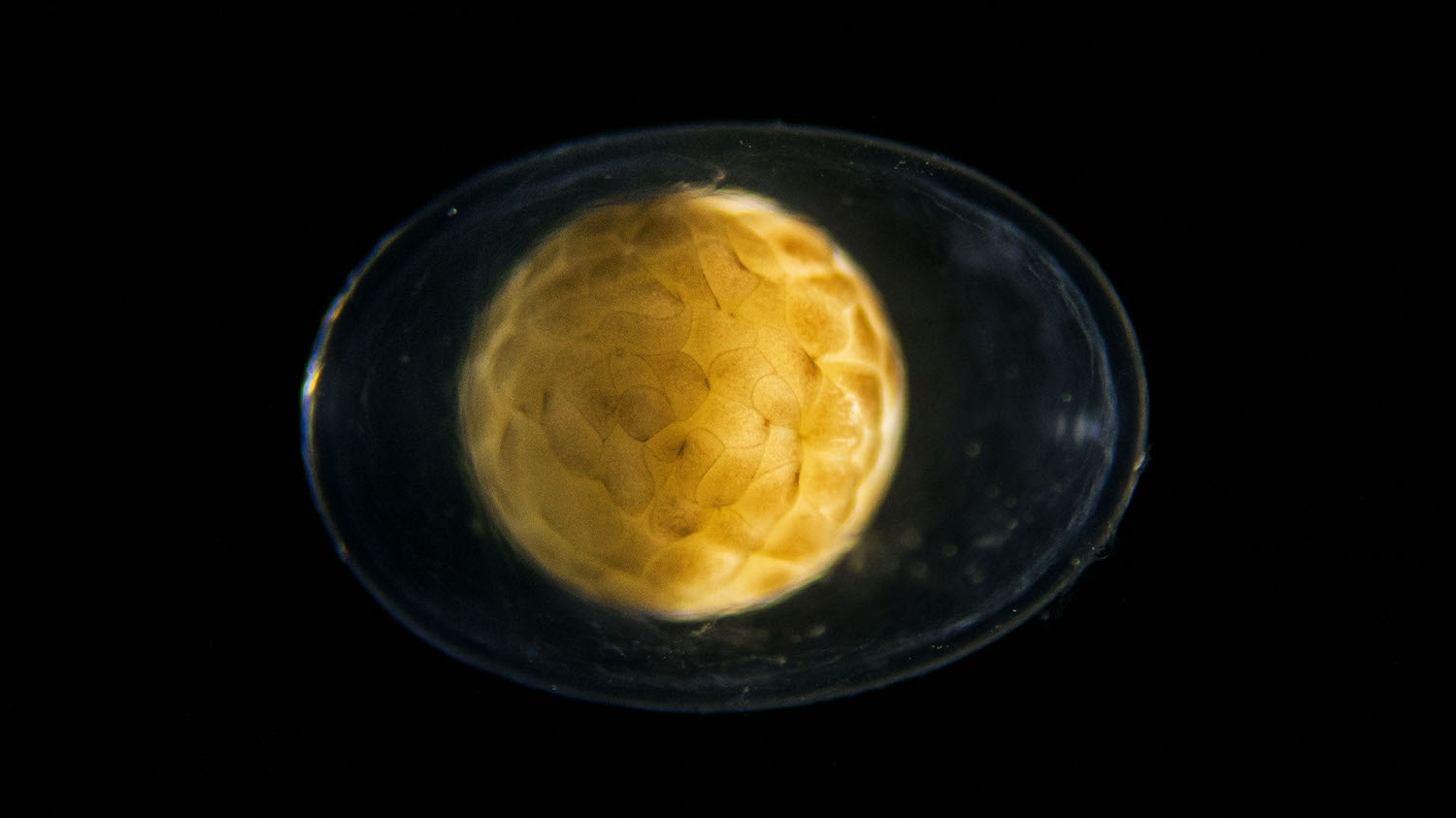
A mesmerizing 6-minute time lapse shows a single cell dividing seemingly endlessly until what was once a yellow blob has become a wriggling, darting salamander tadpole.
"I wanted to film the origin of life," said Jan van IJken, a a photographer and filmmaker based in the Netherlands who created the recently released short, called "Becoming."
But what exactly is happening in this film? Live Science called a developmental biologist to learn more. [Photos: Bizarre Frogs, Lizards and Salamanders]
Van IJken was smart to choose an amphibious alpine newt. "You can look straight into the egg," he told Live Science. "They are transparent, and you can see the whole process." So, he contacted a salamander breeder and picked up a few dozen fertilized eggs.
But there are only a few hours between fertilization and the first cell division, so van IJken had to race home and, like a microsurgeon, unfurl and unglue each egg from the leaf where the salamander mother had carefully stuck it. "Sometimes, I was just in time," van IJken said.
Then, he placed the eggs in water-filled petri dishes and took thousands of photos using a camera attached to a microscope for the next four weeks.
In that initial shot, you can see the fertilized egg (also called an embryo) in the clear, protective vitelline membrane, said Lionel Christiaen, an associate professor of biology at New York University, who wasn't involved with the film. This membrane "helps keep the egg moist and prevents pathogens from getting in," Christiaen told Live Science.
Get the world’s most fascinating discoveries delivered straight to your inbox.
Then, the embryo divides like a maniac. Rather than expanding in size, the embryo increases its number of cells with every division, all within in that same amount of space. There is a lag as each cell replicates the genetic material within it and then divides, in a process called mitosis, Christiaen said.
At about the minute mark in the film, a "hole" appears in the embryo. The process that creates the hole is called gastrulation, when the embryo organizes itself into three distinct cell layers. The importance of gastrulation was captured by Lewis Wolpert, a retired developmental biologist, who famously said, it's "not birth, marriage or death, but gastrulation which is truly the most important time in your life."
At the gastrulation stage, the embryo consists of thousands of cells, and some already "know" that they, or their progeny, will become brain cells, gut cells or something else. "But many of these cells are still on the outside of the egg," Christiaen said. During gastrulation, cells move around, organizing themselves by going to the outermost layer, or ectoderm (nervous system, skin cells and pigment cells); the mesoderm (gut, muscles and red blood cells); or the inner layer, or endoderm (lung cells, thyroid cells and pancreatic cells).
At about the 1:50 mark, the embryo looks like it's putting on a coat. This process is known as neurulation, Christiaen said, and it happens as the neural tube rolls up. After this step, almost everything on the outside of the embryo is there to stay. This consists mostly of the organism's protective skin. [In Photos: How Snake Embryos Grow a Phallus]
At about 3 minutes into the video, you can see the limb buds forming. Soon enough, you can distinguish the head from the tail. Van IJken stopped taking photos for the time lapse and switched to video as soon as the embryo moved, he noted.
Shortly after that, a tube that eventually becomes the heart begins to form, Christiaen said. And once the heart beats, blood flows. You can even see the blood flowing through the gills, the structures that help the animal with gas exchange so that it can breathe underwater.
The developing salamander twitches as it gets older, likely because its growing brain is learning how to control the animal's muscles, Christiaen said.
Finally, the yellow tadpole breaks free from the protective membrane. It's unclear how the animal knows when to do this, but hormones could play a role, Christiaen said. "There's no satisfying answer" to that question, he said.
Watching the tadpoles hatch "was incredible" van IJken said. "How this internal clockwork makes the whole thing come to life, it's incredible. It's a true miracle, one cell dividing and then becoming this animal."
And the circle of life continues, he noted. After the tadpoles hatched, van IJken gave them back to the breeder and got to work editing the film.
Editor's note: You can also see "Becoming" on Jan van IJken's Vimeo page.
- Photos: World's Cutest Baby Wild Animals
- In Photos: Lost Salamanders Discovered
- 7 Ways Pregnant Women Affect Babies
Originally published on Live Science.

Laura is the managing editor at Live Science. She also runs the archaeology section and the Life's Little Mysteries series. Her work has appeared in The New York Times, Scholastic, Popular Science and Spectrum, a site on autism research. She has won multiple awards from the Society of Professional Journalists and the Washington Newspaper Publishers Association for her reporting at a weekly newspaper near Seattle. Laura holds a bachelor's degree in English literature and psychology from Washington University in St. Louis and a master's degree in science writing from NYU.
 Live Science Plus


