Images: Brains with CTE
Get the world’s most fascinating discoveries delivered straight to your inbox.
You are now subscribed
Your newsletter sign-up was successful
Want to add more newsletters?

Delivered Daily
Daily Newsletter
Sign up for the latest discoveries, groundbreaking research and fascinating breakthroughs that impact you and the wider world direct to your inbox.

Once a week
Life's Little Mysteries
Feed your curiosity with an exclusive mystery every week, solved with science and delivered direct to your inbox before it's seen anywhere else.

Once a week
How It Works
Sign up to our free science & technology newsletter for your weekly fix of fascinating articles, quick quizzes, amazing images, and more

Delivered daily
Space.com Newsletter
Breaking space news, the latest updates on rocket launches, skywatching events and more!

Once a month
Watch This Space
Sign up to our monthly entertainment newsletter to keep up with all our coverage of the latest sci-fi and space movies, tv shows, games and books.

Once a week
Night Sky This Week
Discover this week's must-see night sky events, moon phases, and stunning astrophotos. Sign up for our skywatching newsletter and explore the universe with us!
Join the club
Get full access to premium articles, exclusive features and a growing list of member rewards.
What CTE Looks Like in the Brain
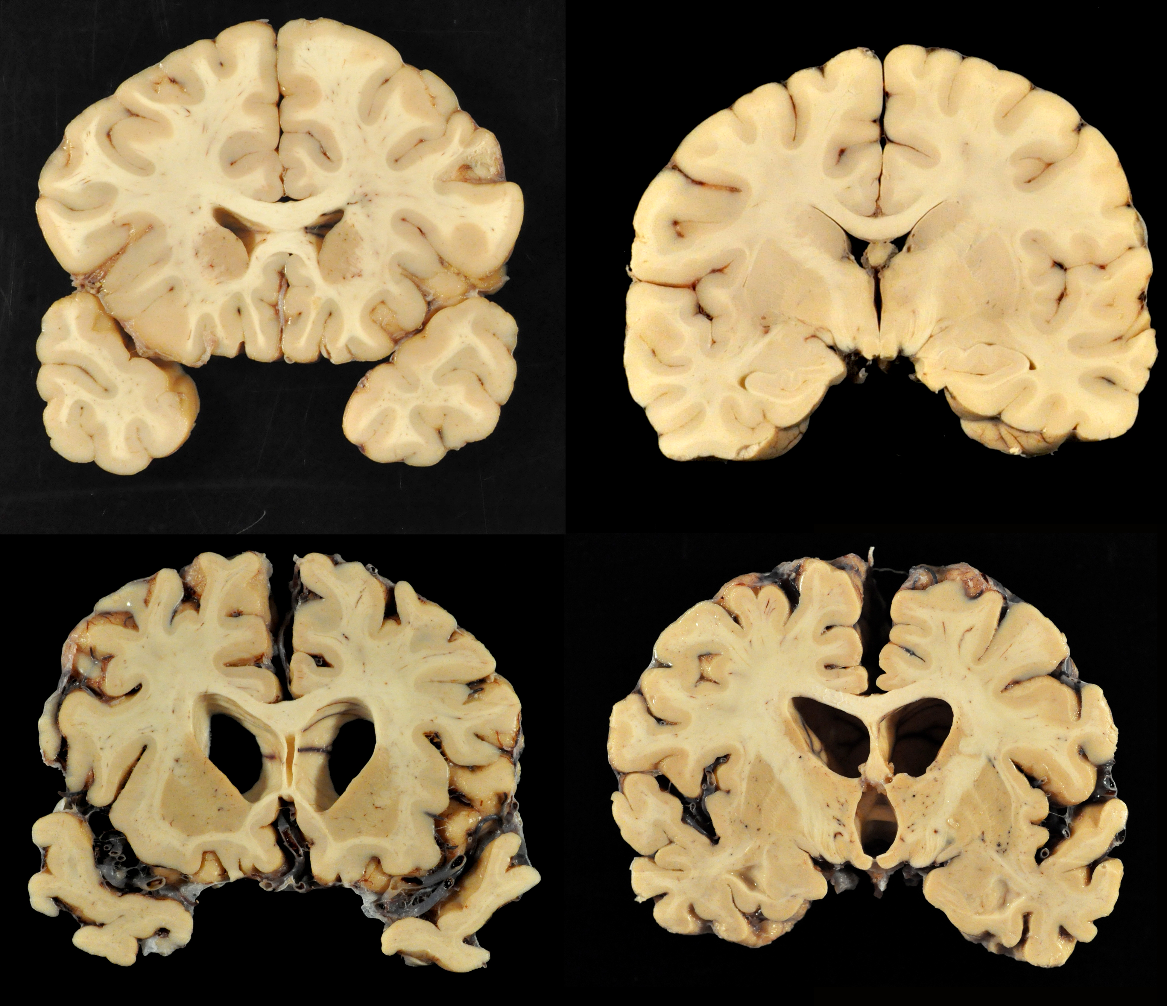
The vast majority of brains donated to science by former football players show signs of the debilitating brain condition chronic traumatic encephalopathy, or CTE, a new report says. [Read our full story on the report.]
CTE is a progressive disease that has been found in athletes such as football players and boxers, who have a history of repetitive blows to the head.
Here, the top two images show a normal brain. The bottom two images show the brain of former University of Texas football player Greg Ploetz, who died with dementia at age 66, in 2015.
Ploetz’s brain revealed that he had severe chronic traumatic encephalopathy, the researchers said. His brain showed atrophy (shrinking), and the brain’s ventricles (the openings in the brain) were larger than normal.
Staining Reveals Tau Protein
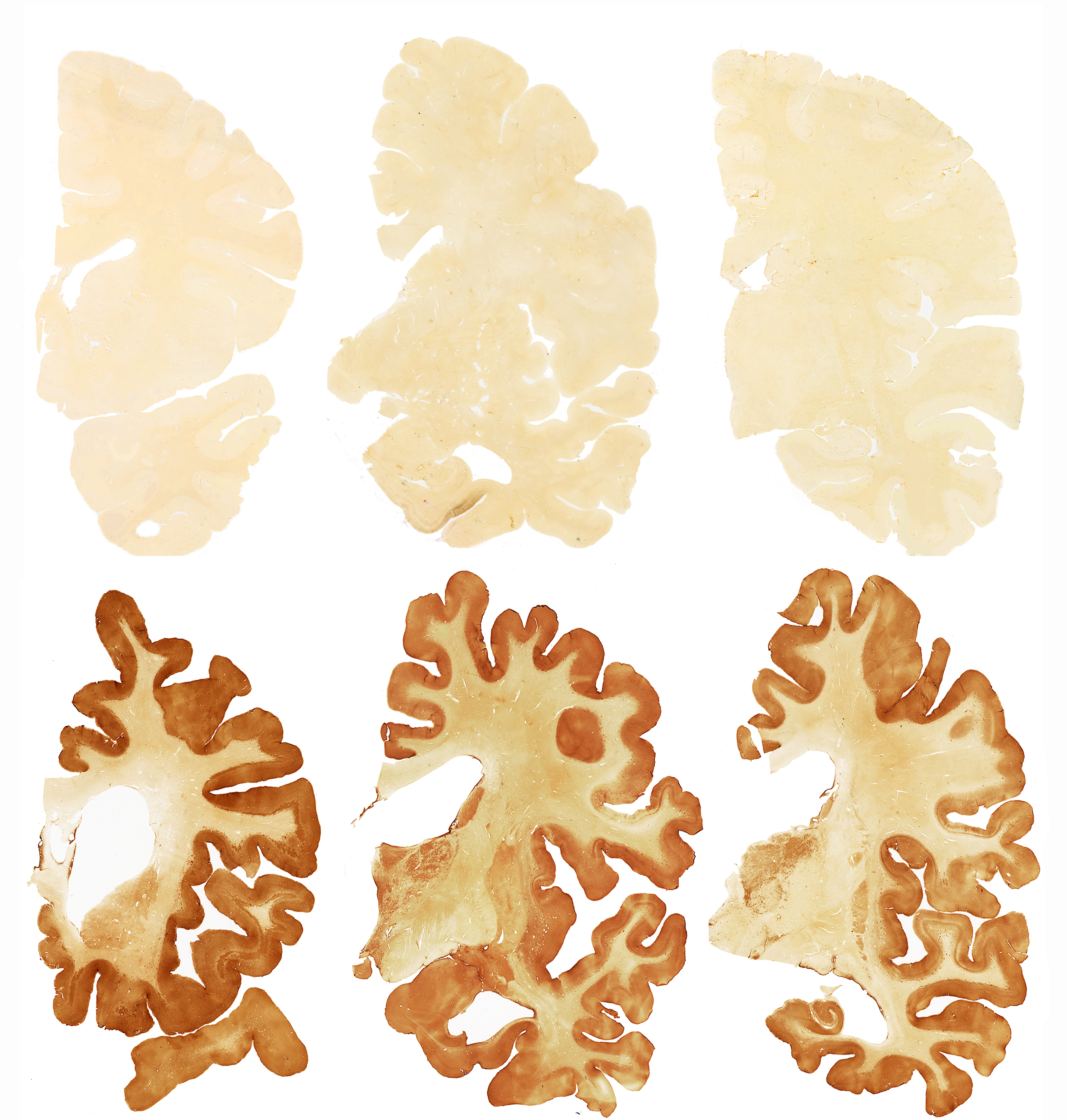
The images in the top row here show a normal brain; the images in the bottom row are of the brain of former football player Greg Ploetz, who had severe CTE. The brown color in Ploetz's brain is the result of a stain that the researchers used to reveal a protein called tau, which is linked with neuron degeneration. Ploetz's brain shows dense tau.
Tau Protein Amid the Brain Cells
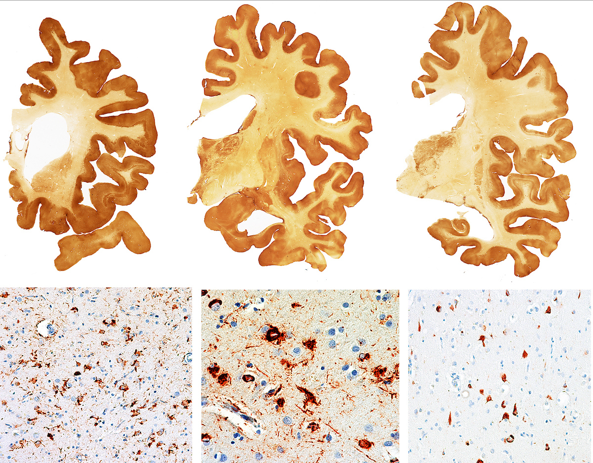
These images show severe CTE in the brain of former University of Texas football player Greg Ploetz. The brown stain reveals the presence of a protein called tau, which is linked with neuron degeneration. The bottom images here show a microscopic view, revealing the dark-stained tau protein amid the neurons and astrocytes (star-shaped cells) of the brain.
Mild and Severe Cases
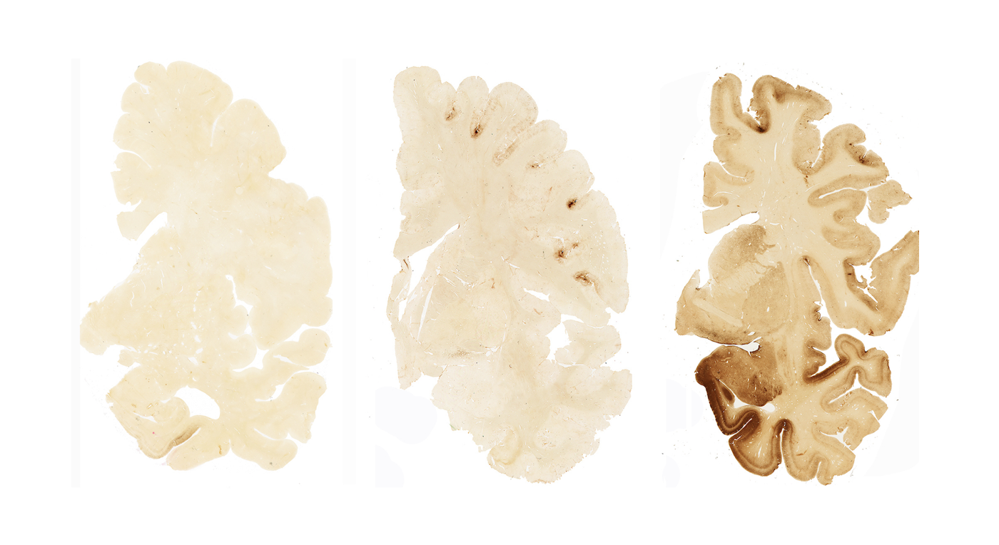
The images here show sections of brain hemispheres. From left to right, the images reveal the differences between a normal brain, the brain of someone with mild CTE, and the brain of someone with severe CTE. The brown stain that was used here reveals the presence of a protein called tau. The normal brain (left) has no obvious signs of tau, the brain of someone with mild CTE (center) shows the presence of some tau, and the brain of someone with severe CTE (right) reveals sections with heavy levels of tau.
Get the world’s most fascinating discoveries delivered straight to your inbox.
Under the Microscope
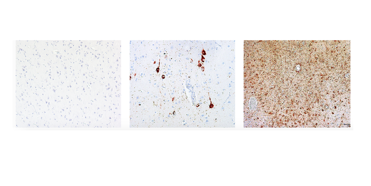
These images show what the brain tissue looks like under the microscope. From left to right, they show a normal brain, the brain of a person with mild CTE, and the brain of a person with severe CTE. The tau protein in the brain is stained, revealing the “tangles” of neuron fibers.
 Live Science Plus


