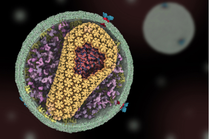
Animating a Cell’s Inner Life
Get the world’s most fascinating discoveries delivered straight to your inbox.
You are now subscribed
Your newsletter sign-up was successful
Want to add more newsletters?

Delivered Daily
Daily Newsletter
Sign up for the latest discoveries, groundbreaking research and fascinating breakthroughs that impact you and the wider world direct to your inbox.

Once a week
Life's Little Mysteries
Feed your curiosity with an exclusive mystery every week, solved with science and delivered direct to your inbox before it's seen anywhere else.

Once a week
How It Works
Sign up to our free science & technology newsletter for your weekly fix of fascinating articles, quick quizzes, amazing images, and more

Delivered daily
Space.com Newsletter
Breaking space news, the latest updates on rocket launches, skywatching events and more!

Once a month
Watch This Space
Sign up to our monthly entertainment newsletter to keep up with all our coverage of the latest sci-fi and space movies, tv shows, games and books.

Once a week
Night Sky This Week
Discover this week's must-see night sky events, moon phases, and stunning astrophotos. Sign up for our skywatching newsletter and explore the universe with us!
Join the club
Get full access to premium articles, exclusive features and a growing list of member rewards.
Animations like this one, which shows HIV infecting a human cell, are helping researchers probe complicated, dynamic molecular interactions.
“In the HIV life cycle, there are a number of events that aren’t really well understood, and people have different ideas of how things happen,” says Janet Iwasa, who made this video by adapting computer programs originally designed to bring characters like Buzz Lightyear to life.
Iwasa is currently working with University of Utah scientists funded by the National Institutes of Health to develop molecularly accurate animations of how HIV enters and exits human immune cells. Animating the stages of viral infection, she says, can give researchers a new way to visualize, communicate and potentially clarify their ideas of how the process works.
When Iwasa was studying cell biology in graduate school, she realized that the only visual representations she had of the proteins she was studying were flat, two-dimensional drawings on paper. She thought, “Why are we relying on oversimplified, static illustrations [of molecules]?”
Within a year, she took an animation class at a local college. She quickly realized that she would need more intensive instruction to be able to animate complex biological processes. A few summers later, she flew to Hollywood for a 3-month training program in industry-standard animation technology.
Iwasa calls her molecular animations “visual hypotheses.” The end results may be beautiful, she explains, but the process of animation itself is what encapsulates and clarifies the science. “In some cases, it might raise more questions and make people go back and do some more experiments when they realize there might be something missing” in their theory of how a molecular process works, she says.
"Janet's animations add great value by helping us consider how complex interactions between viruses and their host cells actually occur in time and space,” said Wes Sundquist, who directs the Center for the Structural Biology of Cellular Host Elements in Egress, Trafficking, and Assembly of HIV (CHEETAH) at the University of Utah. "By showing us how different steps in viral replication must be linked together, the animations suggest hypotheses that hadn’t yet occurred to us. They’re also a lot of fun to watch!"
Get the world’s most fascinating discoveries delivered straight to your inbox.
In addition to HIV infection, Iwasa’s visualizations have helped researchers explore such complex actions as how cells ingest materials, how proteins are transported across a cell membrane and how motor proteins help cells divide. She and her colleagues recently launched Molecular Flipbook, an open-source software toolkit for biologists who want to make their own molecular animations.
This work was funded in part by NIH under grants P50GM082545, RC2GM092708 and R01GM082949.
This Inside Life Science article was provided to Live Science in cooperation with the National Institute of General Medical Sciences, part of the National Institutes of Health.
 Live Science Plus


