
 Live Science Plus


Amazing Look at Fruit Fly Eye (Photo)

By
Nina Sen
published
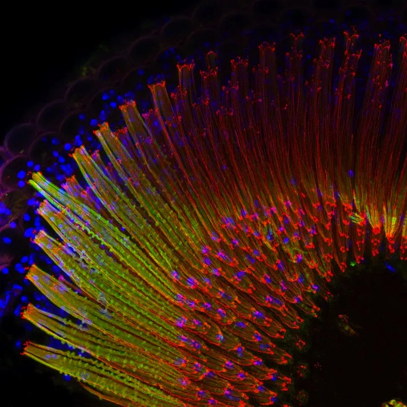
The image represents fruit fly (Drosophila melanogaster) ommatidia, which are arranged in an extremely regular array in the compound eye. Nuclei are shown in blue (DAPI), cadherin in red, and chaoptin in the photoreceptors in green. This Zeiss LSM780 confocal (NA 1.4/40x) image was deconvolved with Huygens Professional.
(Image credit: The Research Institute of Molecular Pathology (IMP))
The spiny globs in this image are part of an eye from a fruit fly (Drosophila melanogaster). Karin Panser from the Research Institute of Molecular Pathology in Vienna received the first prize in the international Huygens Image Contest 2013 for the microphotograph.
The image reveals the imagine units called ommatidia arranged in the compound eye of the fruit fly. Compound eyes contains hundreds to thousands of ommatidia, each equipped with a tiny lens and crystalline cone that transports the light to light-sensitive cells. Compared with simple eyes, compound eyes possess a wide-angle view and are able to detect fast-eye movement.

TOPICS
