Magnificent Microphotography: 50 Tiny Wonders
In a Drop of Water
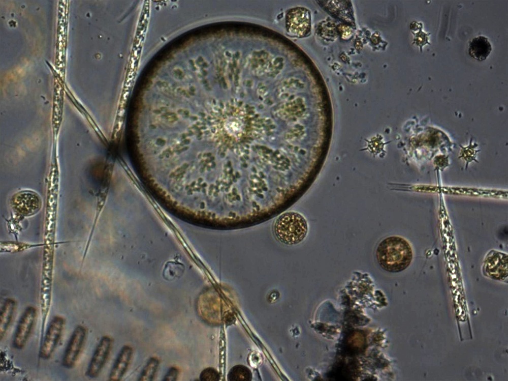
These tiny phytoplankton, called diatoms, are the workhorses of the sea, producing much of the carbon and oxygen in the oceans. A new study in the journal Nature finds that diatoms share at least one molecular process once thought unique to animals, suggesting that the ancestors of diatoms were possibly more closely related to the ancestors of animals than to plants.
Hitch a Ride on a Dragonfly
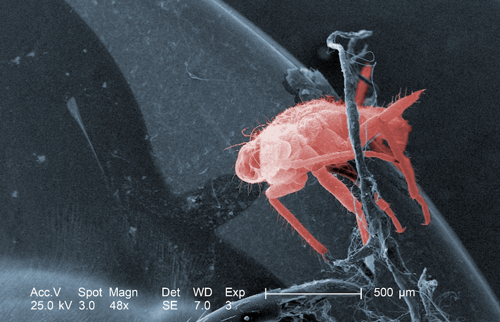
A close-up look at a dead dragonfly found in Georgia revealed this miniature hanger-on. The tiny insect seen in this scanning electron microscope image may have been a dragonfly parasite. Or the bug could be nothing more than debris picked up by the dragonfly on its travels.
Small But Social
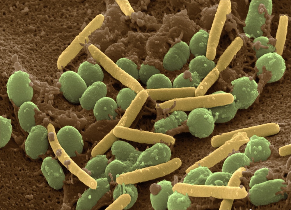
Coming to a clump of dirt near you... Myxococcus xanthus is a social bacterium that preys on other microbes in the soil. When food is abundant, the bacteria take a rod-shaped form, shown here in yellow. When times are tough, bacteria cells clump together into multicellular fruiting bodies containing long-lasting spores, seen here in green.
Some bacteria try to game the system, however, by jockeying to become the hardy spore rather than the supporting fruiting body.
A new study published in the journal Proceedings of the National Academy of Sciences finds that some bacteria in the community evolve to "police" these cheaters, a very primitive form of social cooperation.
It's Not Grandma's Lace
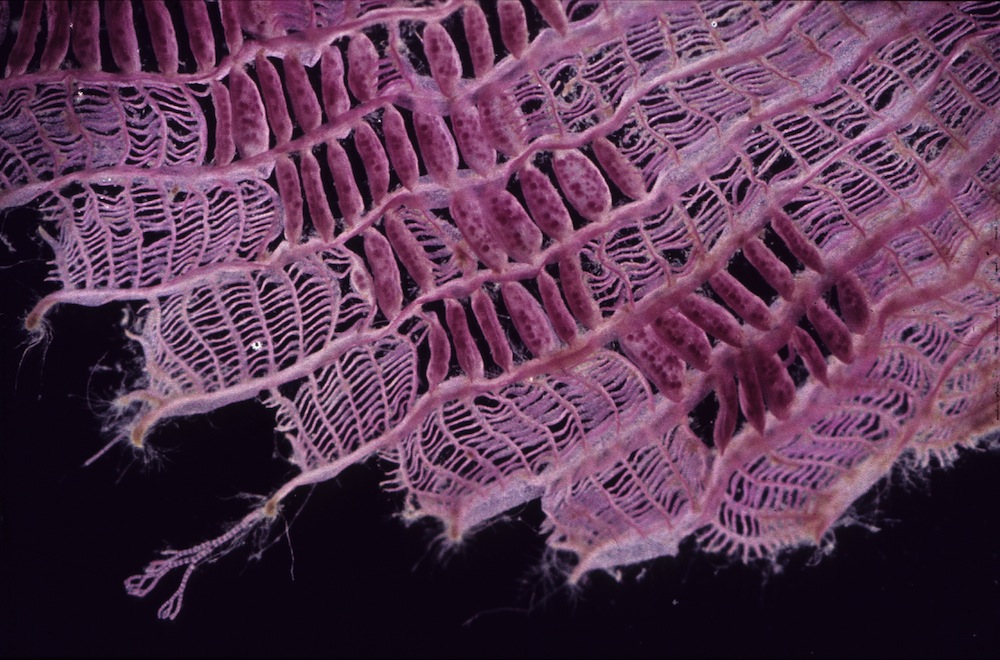
A half-finished crochet project? A tattered scarf? Nope — this is a close-up of Claudea elegans, sea algae found off the coast of Australia.
— Stephanie Pappas
Are We In Outer Space?
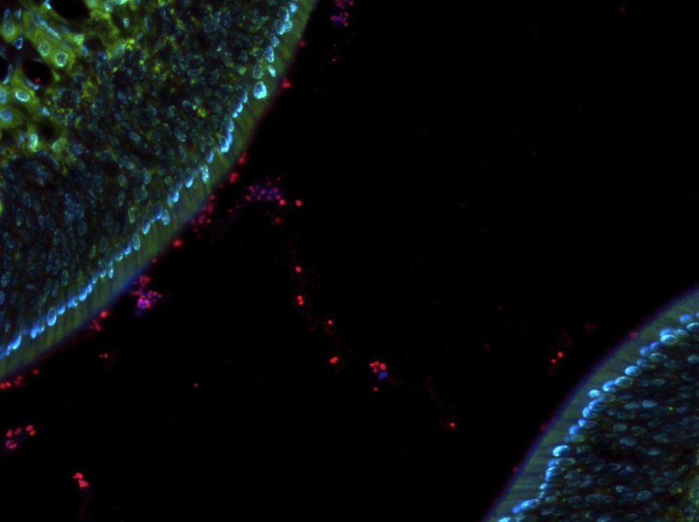
Nope. This is inner space.
The space between cells is a freeway when you're a Staphylococcus bacterium. A tight barrier of cells is supposed to prevent outside invaders like these Staph bugs (red and purple) from entering the body. The fact that we get sick is testimony that those barriers sometimes fail. Now, University of Pennsylvania researchers have found one reason why: Some pathogenic bugs have the key that opens secret passages in this cellular wall.
The surface cells in the respiratory system (shown here in blue) let their guard down when they come in contact with certain pathogen molecules. These molecules trigger the respiratory cells to stop producing proteins that keep the junctions between cells tight. Once that happens, it's no problem for the tiny, deadly microbes to breeze through like they own the place.
— Stephanie Pappas
Who's Doing the Wave?
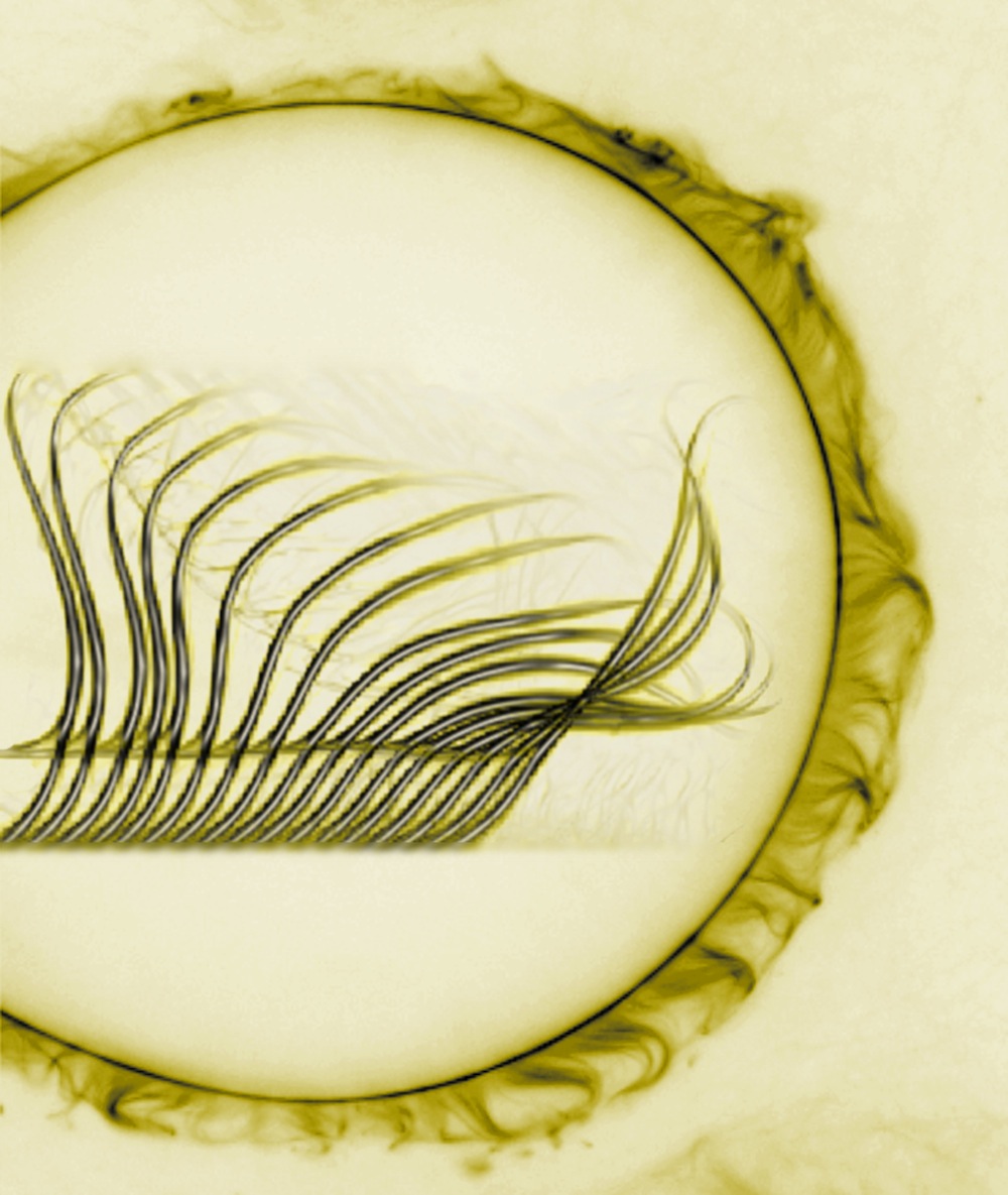
Here's a hint: Something really small.
These are a laboratory-built version of cilia, tiny hair-like projections off of a cell body. In a cell, cilia beat in synchronization much like "The Wave" so beloved by sports fans, propelling a cell or brushing away foreign material (cilia in our lungs help expel inhaled particles, for example.)
Using just four cellular components, researchers at Brandeis University in Massachusetts found that they could build super-simple cilia that automatically sync up with one another, beating in perfect rhythm. We'd like to see a bunch of drunk baseball fans manage that.
— Stephanie Pappas
Tiny Feet Take Big Steps for Cancer Cells
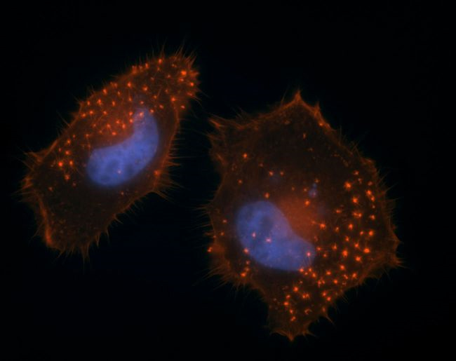
The spread of cancer from one its initial outpost to someplace else in the body, called metastasis, is the most common reason cancer treatments fail. Some cancer cells rely on microscopic "feet" called invadopodia, which are projections on the cellular membrane that help the cells "walk" to surrounding tissues. Now researchers are reporting online in the July 26, 2011, issue of the journal Science Signaling that they have identified compounds that inhibit invadopodia formation without causing toxicity. The team also found a number of compounds that increased a cancer cell's invadopodia.
Get the world’s most fascinating discoveries delivered straight to your inbox.
Here, invadopodia (bright red dots) form on metastatic cancer cells.
The Forest in Your Eye
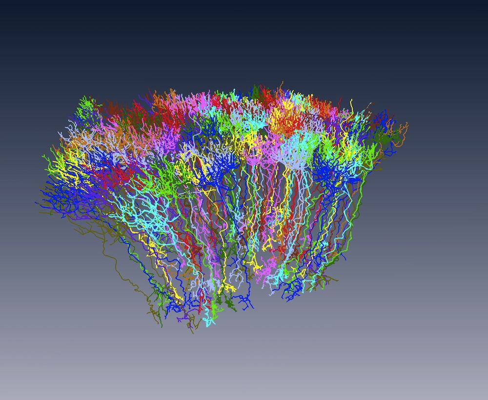
These candy-colored "trees" are actually the cells that enable you to see in the dark. They're called rod cells, and humans have some 120 million of them lining the back of the eye, shooting signals to the brain when they're stimulated by light. Rods are sensitive to very dim light, unlike their counterparts, cones, which allow us to see color.
Scientists at the Max Planck Institute for Medical Research in Heidelberg made this image using new brain-mapping software that traces the connections between nerve cells 50 times faster than earlier methods. The process has now been tested on the mouse retina, as seen above, and researchers plan to tackle the rodent's cerebral cortex next. For more amazing brain images, check out LiveScience's gallery, Inside the Brain: A Journey Through Time.
—Stephanie Pappas
How Do Your Guts Grow?
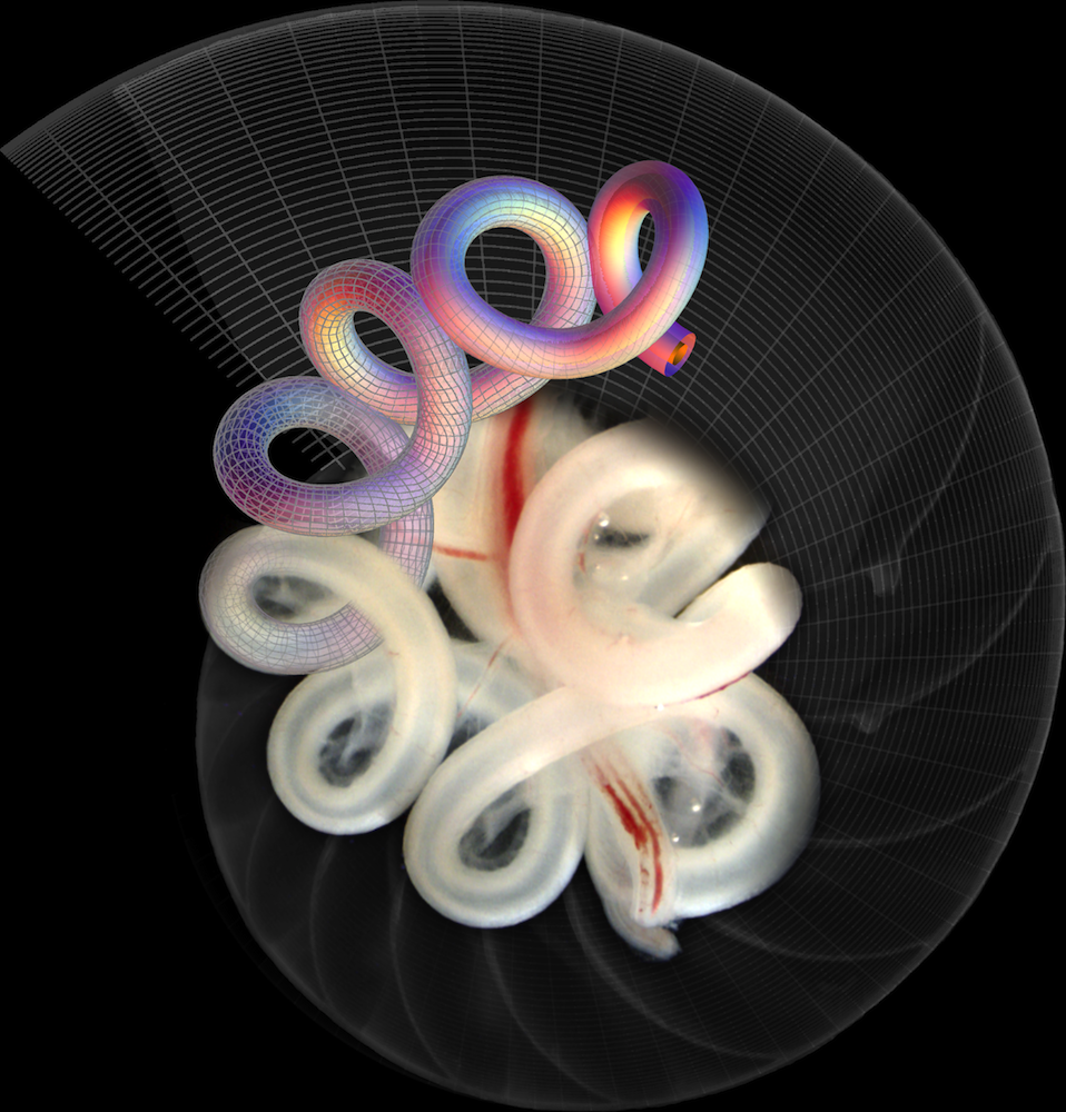
As fetal you developed in the womb, your intestines grew faster than your body, forcing the guts to loop around on themselves. A new study published Aug 4 in the journal Nature found that the patterns of this fold depend on the elasticity, geometry and rate of growth of the gut and the muscles it's anchored to.
Here, a chick's gut melds with a numerical simulation of chicken gut development.
— Stephanie Pappas
Eggshells Hold Hidden Worlds
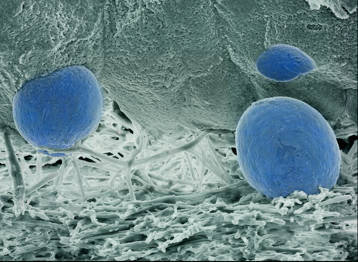
This image taken by Hanna Jackowiak shows the microstructures of the lower parts of eggshell wall in a pheasant. The eggshell in birds is composed of a thick layer of mineral column and underlying thin, fibrous membrane. Scanning electron microscopy was used to show the space between these layers.
This image was taken during microscopic studies on the spatial structure of the eggshell in the pheasant and was an entry in the 2005 Science & Engineering Visualization Challenge (SciVis) competition, sponsored by the National Science Foundation and the Journal Science. The competition is held each year to recognize outstanding achievements by scientists, engineers, visualization specialists and artists who are innovators in using visual media to promote the understanding of research results and scientific phenomena. To learn more about the competition and view all the winning entries, see the SciVis Special Report. (Date of Image: May 30, 2005.)

Stephanie Pappas is a contributing writer for Live Science, covering topics ranging from geoscience to archaeology to the human brain and behavior. She was previously a senior writer for Live Science but is now a freelancer based in Denver, Colorado, and regularly contributes to Scientific American and The Monitor, the monthly magazine of the American Psychological Association. Stephanie received a bachelor's degree in psychology from the University of South Carolina and a graduate certificate in science communication from the University of California, Santa Cruz.


